In Vivo Measurement of Hindlimb Dorsiflexor Isometric Torque from Pig
The present protocol describes concise experimental details on the evaluation and interpretation of in vivo torque data obtained via electrical stimulation of the common peroneal nerve in ...
Out of This World Research at Brock University: Space Flight’s Impact on Female Mice Reproductive Health
NASA's SpaceX CRS-29 launched on November 9th carrying the Rodent Research-20 (RR-20) payload, the contents of which will be studied by Brock University's Dr. Val Fajardo and his team using Aurora ...
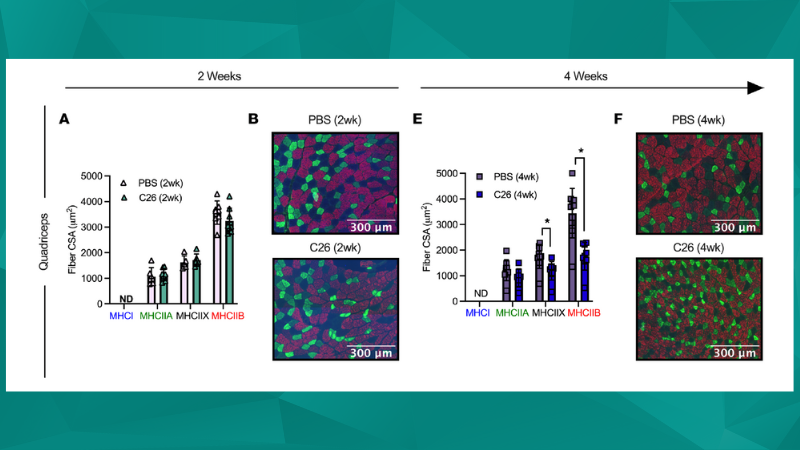
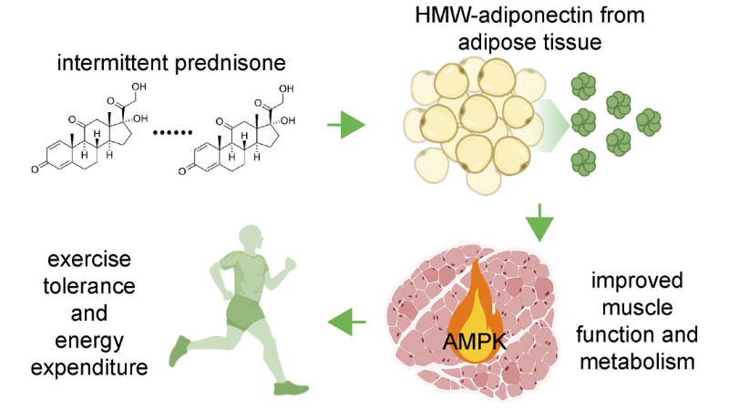
Cancer Cachexia-Induced Muscle Atrophy
Cancer cachexia is a muscle wasting syndrome that is associated with certain cancers, but most commonly with advanced malignancies. This syndrome arises as a result of tumor-induced metabolic ...
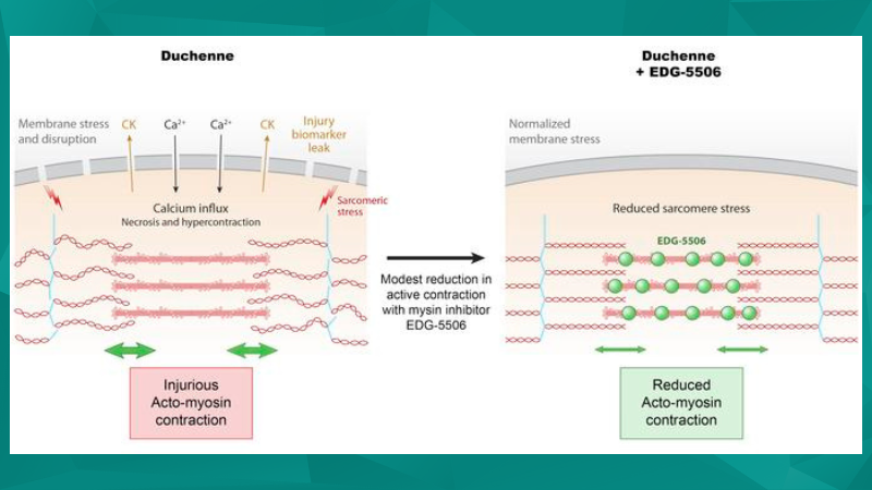
Recent Therapeutic Advances in Duchenne Muscular Dystrophy (DMD) Models
The following publication review showcases recent studies focused on therapeutic advances in Duchenne Muscular Dystrophy (DMD) that use Aurora Scientific ...
Exercise Science
Aurora Scientific equipment has played important roles in helping researchers understand muscle physiology under a variety of conditions in animal models as highlighted in this publication ...
Designing an in-vivo study in DMC LabBook
This blog will provide a walkthrough of how to design a study in our Dynamic Muscle Control (DMC) LabBook software, specifically for 1300A 3-in-1 Whole Animal System in-vivo experiments.
Excellence, Innovation, and Collaboration: Key Research at the MHRC
This past April, we partnered with InsideScientific and the Muscle Health Research Centre (MHRC) at York University, a one-of-a-kind facility that fosters an interdisciplinary approach to the study ...
Sensory Encoding by Muscle Spindle Afferents
This publication review highlights Aurora Scientific instruments that can facilitate the study of sensory encoding by muscle spindle ...
Excellence, Innovation, and Collaboration: Student Research at the MHRC
This past April, we partnered with InsideScientific and the Muscle Health Research Centre (MHRC) at York University, a one-of-a-kind facility that fosters an interdisciplinary approach to the study ...
Functional Recovery of the Musculoskeletal System Following Injury – Leveraging the Large Animal Model
Watch Dr. Sarah Greising discuss the current pathophysiologic understanding of the skeletal muscle remaining following traumatic musculoskeletal ...
Excellence, Innovation, and Collaboration: A Day at the MHRC with Arthur Cheng
Interviewing Dr. Arthur Cheng at the Muscle Health Research Centre (MHRC), York University, an innovative research centre that facilitates the interdisciplinary study of muscle biology and the ...