New Year, New Insights: Unique Models in Muscle Physiology
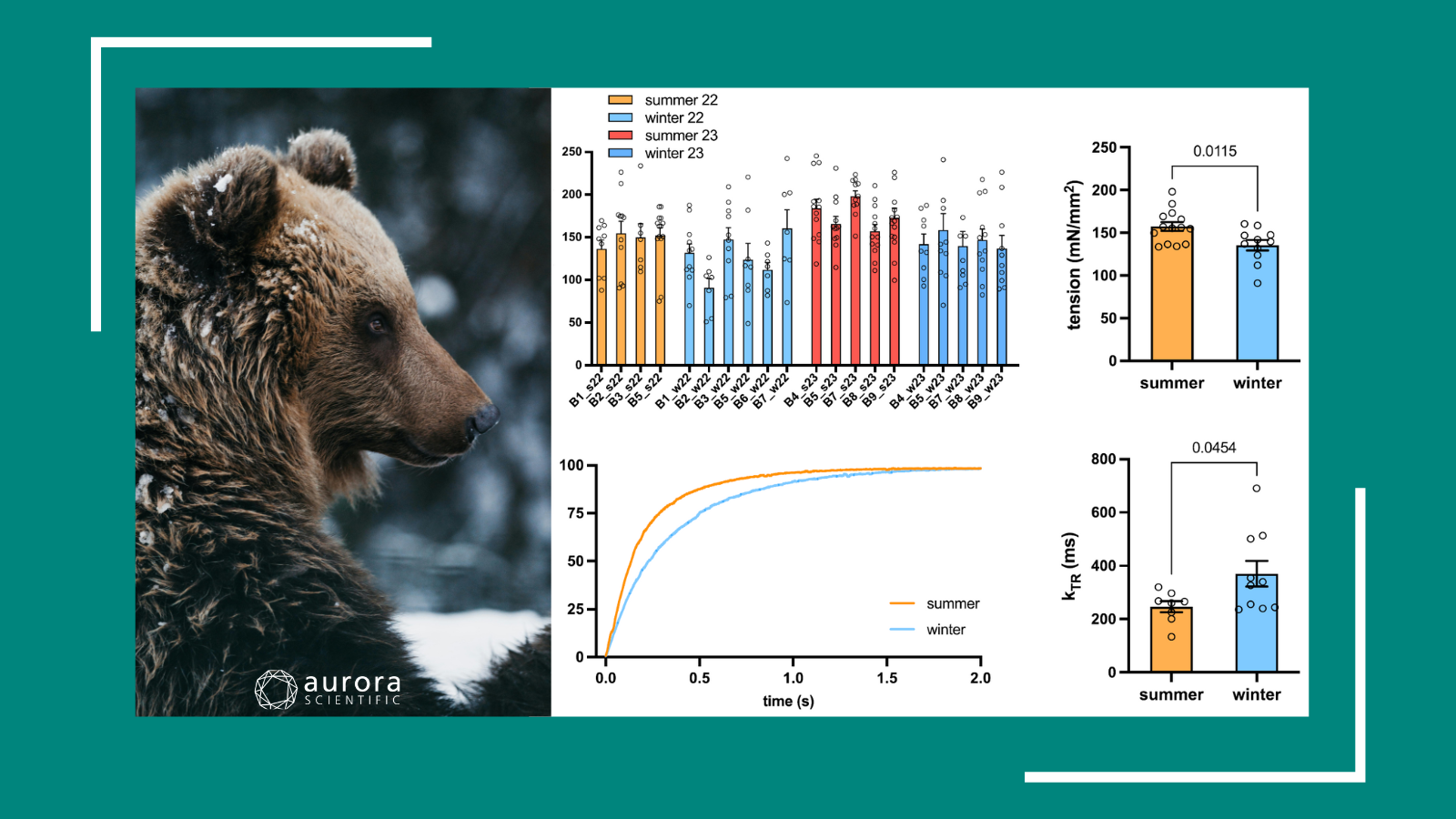
Ringing in the new year is an exciting time for resolutions, recalibrations, and research. The ‘New Year, New Me’ mindset can help kickstart a period of personal growth, and recent research ...

Writing Protocols with 600A for Permeabilized Tissues
This blog will provide a brief overview of how to write protocols using our Real-Time Muscle Data Acquisition and Analysis System (600A) software.

2025 Winter/Spring Scientific Conferences and Meetings
The Aurora Scientific team is continuing to journey out and connect with researchers at scientific conferences and meetings all over the world this Winter and Spring! We are particularly thrilled to ...
Molecular Signals Mediating Increases in Muscle Size and Function
In this webinar, Dr. Bert Blaauw elucidates skeletal muscle regulatory pathways and offers approaches to tackle muscle deficits for ...

Worth the Weight: Impacts of Microgravity on Muscle Health
In anticipation of the 2024 American Society for Gravitational and Space Research (ASGSR) conference, the following publication launches into the latest insights on microgravity and muscle health.

Quick Start Guide to 600A
This blog will provide a brief overview of how to start-up and utilize our Real-Time Muscle Data Acquisition and Analysis System (600A) software.
Down to the Muscle: Sarcopenia Modelling and Interventions
Over the years, research into sarcopenia and aging has increasingly focused on improved models, mechanistic insights, and intervention testing. To highlight this trend, the following review covers ...
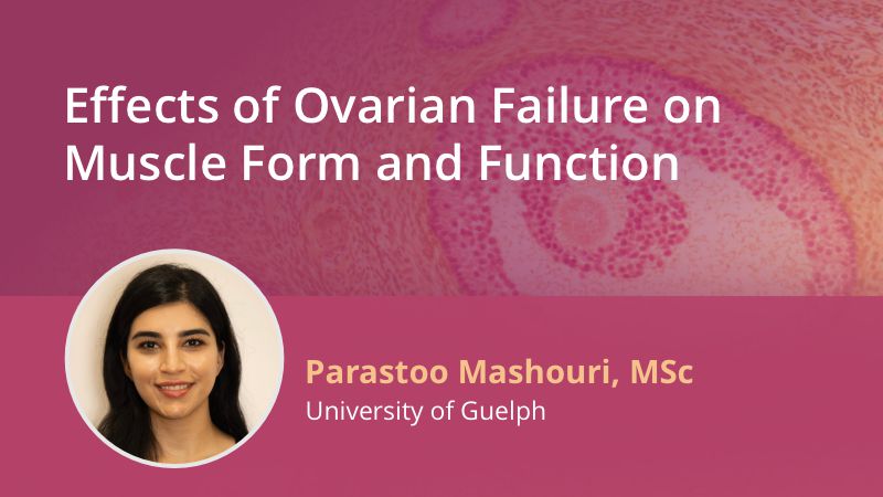
WEBINAR: Effects of Ovarian Failure on Muscle Form and Function
Live October 30th, this presentation features Parastoo Mashouri highlighting her pioneering research on the effects of gradual ovarian failure on skeletal muscle ...
Out of This World Research at Brock University: Post Mission Insights into Alterations of Soleus Muscle Function in Space-Flown Mice
Hear Dr. Val Fajardo, Jessica Braun, and Briana Hockey on their experience working on NASA's Rodent Research-20 (RR-20) mission at the Roskamp Institute, the data collected utilizing Aurora ...

Step-by-Step: Analyzing Experiments in DMA
This blog will provide a walkthrough of how to analyze an experiment in our Dynamic Muscle Analysis (DMA) LabBook ...
Moving Mountains: Recent Feats in Muscle Physiology
As we March towards the 2024 American Physiology Summit, this month’s publication review covers recent advancements in the realm of muscle physiology, including the development of an improved ...
2024 Spring/Summer Scientific Conferences and Meetings
The Aurora Scientific team is continuing to journey out and connect with researchers at scientific conferences and meetings all over the world this spring and summer! We are particularly thrilled to ...
MYTHO is a novel regulator of skeletal muscle autophagy and integrity
The study identifies a novel FoxO-dependent gene called Mytho (Macroautophagy and YouTH Optimizer) as a regulator of autophagy and skeletal
Muscle weakness precedes atrophy during cancer cachexia and is linked to muscle-specific mitochondrial stress
Cancer-induced cachexia is a complex syndrome marked by skeletal muscle mass loss, impacting functional independence, quality of life, and cancer treatment outcomes. While muscle wasting has traditionally been attributed to circulating factors, recent research revealing mitochondrial dysfunction preceding the onset of muscle atrophy suggests it may play a role. Understanding the muscle-specific and time-dependent nature of mitochondrial responses to cancer may offer insights for targeted therapeutic interventions to mitigate muscle weakness and wasting in cachexia. This study examines the relationship between muscle dysfunction and mitochondrial bioenergetics in locomotor and respiratory muscles, revealing diverse responses that underscore the need for tailored approaches to treating muscle complications in cancer cachexia.
Blocking muscle wasting via deletion of the muscle-specific E3 ligase MuRF1 impedes pancreatic tumor growth
Cancer-induced muscle wasting significantly impacts quality of life, hinders cancer treatments, and predicts early mortality. This study explores the role
Microdystrophin Gene Addition Significantly Improves Muscle Functionality and Diaphragm Muscle Histopathology in a Fibrotic Mouse Model of Duchenne Muscular Dystrophy
This study investigates the effectiveness of adeno-associated viral (AAV) vector-mediated microdystrophin gene addition therapy in a fibrotic mouse model of