The field of exercise science encompasses a broad range of study, for which Aurora Scientific has a number of solutions. This publication review highlights how some of our instruments have been used to study the morphological characteristics and function of muscle, as well as myocellular signaling, in rodent models of exercise.
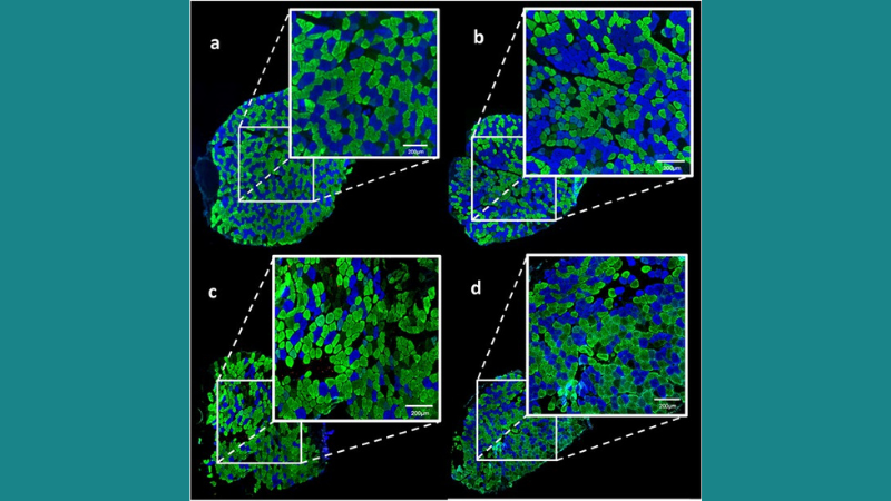
Featured image demonstrates that low-intensity eccentric training (LIET) improves muscle fiber distribution in mdx mice (© 2021 Pedrazzani, P. S. et al.).
Twenty-One Days of Low-Intensity Eccentric Training Improve Morphological Characteristics and Function of Soleus Muscles of mdx Mice
Duchenne muscular dystrophy (DMD) is a muscle disorder that leads to muscle weakness, progressive degeneration, and cardiac and respiratory failure. While there is no cure for DMD, rehabilitation exercises can slow disease progression. Eccentric training can benefit disused muscles, but no studies to date have examined how LIET affects dystrophic muscles. Therefore, Pedrazzani et al. investigated the effects of LIET on dystrophic soleus muscle in mdx mice, a common animal model for DMD. Following training, muscle fragments of euthanized mice were removed for various analyses, including single fiber force measurements using our 403A force transducer, 322C length controller and 802D permeabilized fiber apparatus. Importantly, 21 days of LIET was shown to significantly increase total force in mdx muscle fibers. Immunofluorescence and histological analyses also revealed that LIET reduces cell degeneration and improves trophism and fiber distribution in mouse dystrophic soleus muscle. The authors concluded that LIET has potential for rehabilitating dystrophic muscle for DMD treatment.
Skeletal Muscle Phenotype Signaling with Ex Vivo Endurance-Type Dynamic Contractions in Rat Muscle
Elucidating the underlying mechanisms of exercise-induced skeletal muscle phenotype transitions aid in understanding skeletal muscle adaptive potential, health, and performance overall. However, most studies to date employ isometric contraction models, which are not representative of the dynamic muscle actions that occur during in-vivo exercise. Therefore, Jakobsgaard et al. employed an ex-vivo dynamic stretch-shortening contraction model to study how skeletal muscle phenotype affects myocellular protein signaling, with a focus on rat soleus and extensor digitorum longus (EDL) skeletal muscle. Force measurements were performed with our 305C dual-mode lever, which allowed for length and tension manipulation of suspended muscle-tendon unit preparations. While modest force development changes were observed in both phenotypes, soleus muscle exhibited greater peak force than EDL muscle due to differences in contractile properties, which could explain the greater p70 S6K phosphorylation observed in soleus muscle. The recognizable signaling response patterns found in this study suggest that this model can resemble in-vivo endurance exercise. The authors concluded that these findings could help interpret myocellular signaling outcomes from mixed muscle samples collected during clinical trials.


