
Exercise Science: A Force of Nature
With our attendance at the 20th International Biochemistry of Exercise Conference (IBEC), we’re excited to spotlight the latest in exercise research. From volumetric muscle loss and dystrophic ...

Springing Forward: Myofilament Advances
From unraveling the roles of titin’s P-zone domains in thick filament arrangement to examining the impact of cMyBP-C mutations on cardiac muscle relaxation, these papers illuminate ongoing efforts ...
What’s New in DMC LabBook v6.1: A Streamlined Workflow for Muscle Researchers
This blog provides an overview of the updates that have been made to the newly unveiled Dynamic Muscle Control LabBook Software, v6.1.
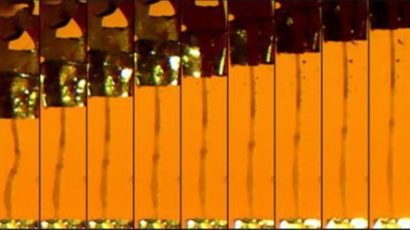
Construction of Constant-Load (Isotonic) and Constant-Velocity (Isokinetic) Torque-Velocity-Power Profiles In vivo for the Rat Plantar Flexors
Quantification of knee extensor maximal strength is imperative to understand functional adaptations to aging, disease, injury, and rehabilitation. We present a novel method to repeatedly measure in ...
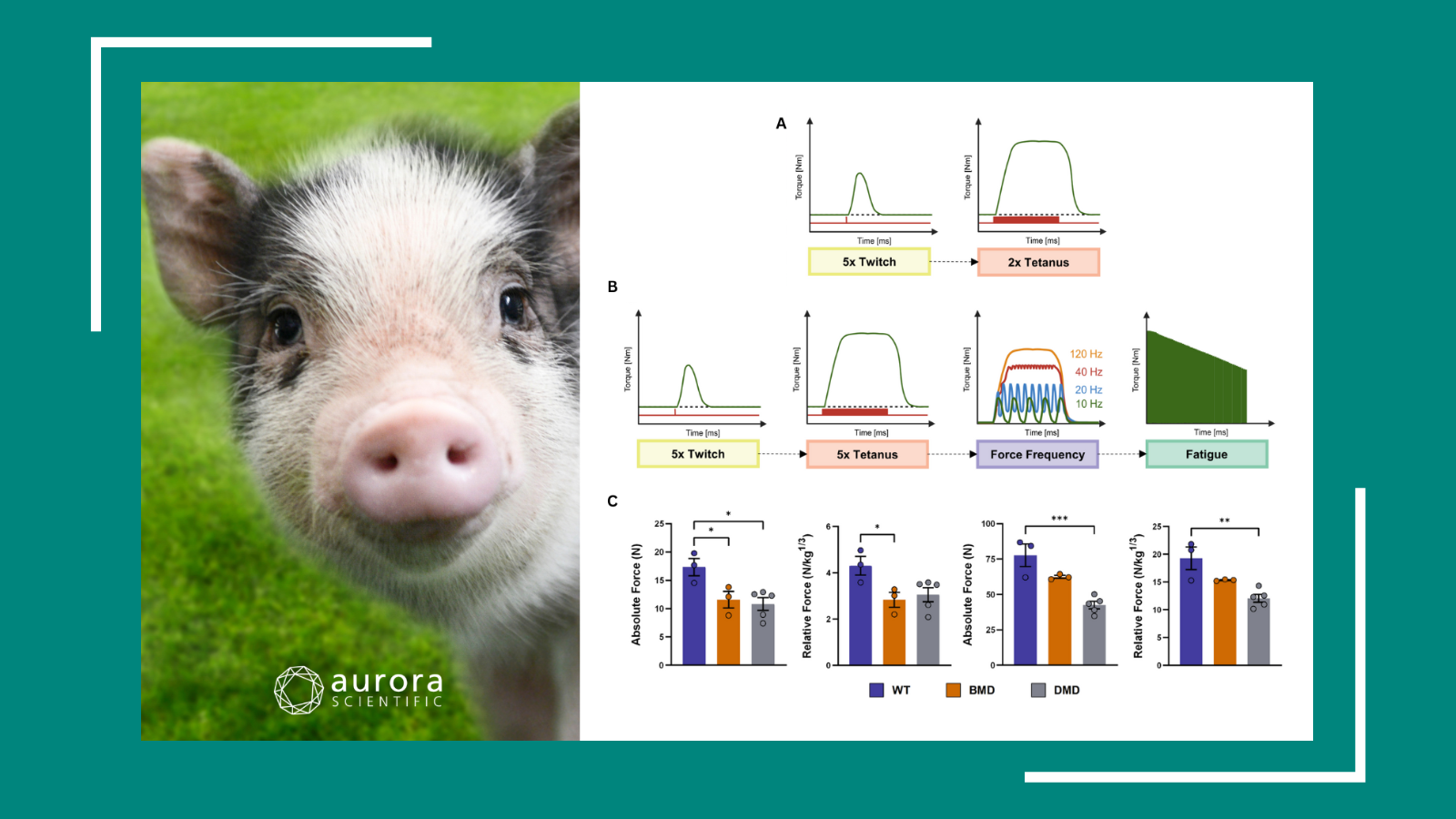
Pig-ture Perfect Progress
Kicking off 2026 with a lucky trot, this publication review highlights cutting-edge porcine research revealing how pigs help unravel the mechanics of muscle and heart function, from dystrophic rescue ...
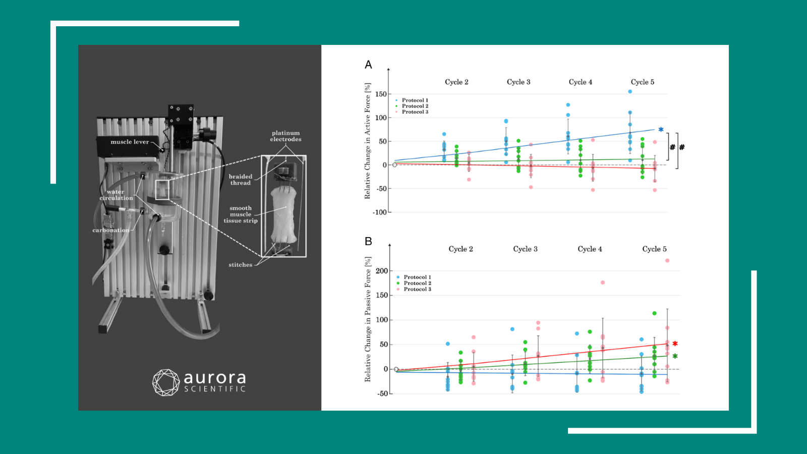
Fueling Function: Forthcoming Insights in Muscle Physiology
In the spirit of the American Physiology Summit, the following publication review covers forthcoming insights in muscle physiology, including smooth muscle mechanics, a neutralizing treatment for ...
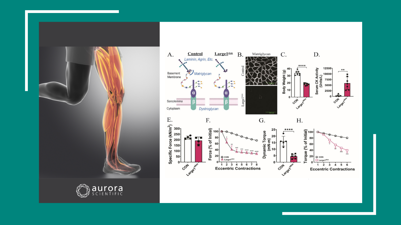
Marching Forward: Recent Skeletal Muscle Discoveries
In conjunction with the Advances in Skeletal Muscle Biology conference, the following publication review digs into the impacts of protein dysfunctions, knockdowns, and modifications on skeletal ...
Writing Protocols with 600A for Permeabilized Tissues
This blog will provide a brief overview of how to write protocols using our Real-Time Muscle Data Acquisition and Analysis System (600A) software.
Quick Start Guide to 600A
This blog will provide a brief overview of how to start-up and utilize our Real-Time Muscle Data Acquisition and Analysis System (600A) software.
Women’s Health Month: Strides in Muscle Physiology
In honour of Women's Health Month, May’s publication review covers recent advances in female-focused muscle physiology research. These investigations into gradual ovarian failure, the impact of ...

Step-by-Step: Analyzing Experiments in DMA
This blog will provide a walkthrough of how to analyze an experiment in our Dynamic Muscle Analysis (DMA) LabBook ...
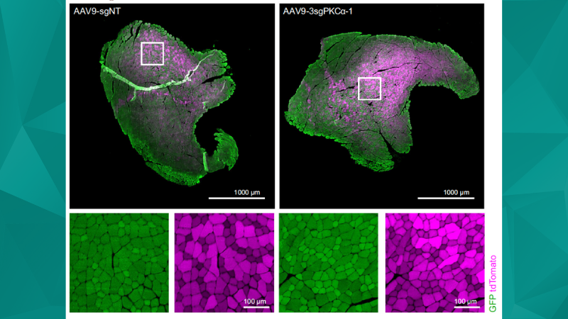
Fast, multiplexable and efficient somatic gene deletions in adult mouse skeletal muscle fibers using AAV-CRISPR/Cas9
Thürkauf et al. establish an efficient CRISPR/Cas9 genome editing system for targeted, multiplexable gene knockouts in skeletal muscle fibers of
Best of 2023: Across Countries and Applications
2023 has proved to be a particularly fruitful year for scientific discovery, with a multitude of pioneering studies spanning continents and disciplines. From the intricate workings of muscle ...

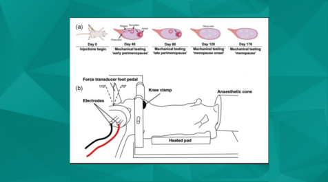
In Vivo Measurement of Hindlimb Dorsiflexor Isometric Torque from Pig
The present protocol describes concise experimental details on the evaluation and interpretation of in vivo torque data obtained via electrical stimulation of the common peroneal nerve in ...
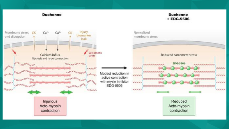
Recent Therapeutic Advances in Duchenne Muscular Dystrophy (DMD) Models
The following publication review showcases recent studies focused on therapeutic advances in Duchenne Muscular Dystrophy (DMD) that use Aurora Scientific ...
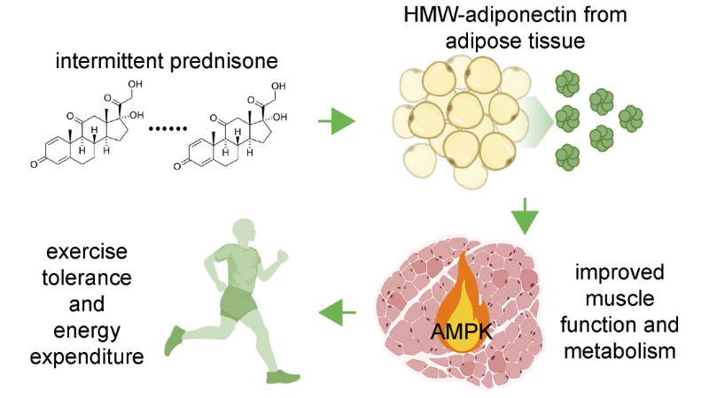
Exercise Science
Aurora Scientific equipment has played important roles in helping researchers understand muscle physiology under a variety of conditions in animal models as highlighted in this publication ...

Neuromuscular Junction and Muscle Pathophysiology
Thanks to Aurora Scientific equipment, numerous groups have been able to further expand our understanding of these pathologies and how we can improve muscle function. This publication review ...
Designing an in-vivo study in DMC LabBook
This blog will provide a walkthrough of how to design a study in our Dynamic Muscle Control (DMC) LabBook software, specifically for 1300A 3-in-1 Whole Animal System in-vivo experiments.
Unique and Interesting Animal Models
At Aurora Scientific, we provide instrumentation to help assess muscle biomechanics in a variety of animal models from flies to octopuses, and highlight some recent examples in this publication ...