Exercise and performance are inextricably linked to the cellular mechanisms and physiology of the muscle. From mitochondria and oxidative metabolism to generated-force, architecture, and tolerance of skeletal muscle, Aurora Scientific equipment has played important roles in helping researchers understand muscle physiology under a variety of conditions in animal models. This publication review highlights the important research that rely on our equipment and software.
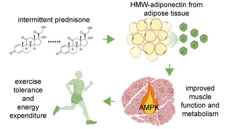
Featured image (©Quattrocelli et al. 2022, licensed under CC BY), is a graphical abstract of the link between intermittent prednisone and the rise in muscle function and metabolism through adiponectin.
Innately Expressed Estrogen-Related Receptors in the Skeletal Muscle are Indispensable for Exercise Fitness
Among the most closely observed lines of research in exercise science, mitochondrial function is integral to exercise performance. The aerobic ability of skeletal muscle is dependent on mitochondrial oxidative capacity and metabolic function. In this article, Sopariwala et al. (2022) sought to determine how two nuclear receptors, estrogen-related receptor alpha and gamma (ERRα/γ), work to regulate exercise capacity. These receptors are among a family of nuclear receptors that are under investigation in regard to their roles in metabolic regulation. They control the metabolics surrounding glucose and fatty acids as well as thermogenesis. The authors’ study aimed to learn more about the ERRs’ contribution to aerobic function, contractility and endurance.
Using single and double knockout mice for the ERRs, the authors observed the impact of the loss of function of one or both of the ERRs on mouse muscle structure and function. Using Aurora Scientific’s 1300A 3-in-1 Whole Animal System, the authors determined the extent of in vivo muscle contractility. The contractile events were recorded using our Dynamic Muscle Control software and calculated with our Dynamic Muscle Analysis software. The single knockout of a either ERRα or ERRγ did not have a significant effect on the muscle, however double knockout of both ERRs cause the muscles to become pale. Sequencing found that the muscle had a decrease in gene expression for oxidative metabolism versus wildtype muscle. Also mitochondrial number, size, and function all declined in the double knockout. Their results show the critical role that ERRα/γ play in muscle function and aerobic capacity.
Intermittent Prednisone Treatment in Mice Promotes Exercise Tolerance in Obesity Through Adiponectin
Circulating signals that regulate metabolic function are abundant in the body. Fat and muscle cross-talk involves one such signal known as adiponectin, which is one of the most abundant adipokine and a key regulator. In obesity, this cross-talk is impaired with little known research into its restoration in muscle. Glucocorticoid steroids such as prednisone when taken chronically leads to obesity and metabolic stress, however, an intermittent dosing was previously found to improve muscle function in muscular dystrophy. The authors of this article, Quattrocelli et al. (2022), believe that the benefit of prednisone could also apply to obesity as it does with muscular dystrophy. They reasoned that adiponectin and the fat-muscle cross-talk can be improved alleviating metabolic stress and exercise intolerance through prednisone treatment, though the mechanisms of this are not clear.
The authors used mice with diet-induced obesity. They utilized an intermittent once-a-week prednisone regimen and observed the changes in exercise tolerance and muscle function. Using our 1300A 3-in-1 Whole Animal System and our 300C-LR Dual-Mode Lever system, in-situ tetanic force was measured in mice. They found that the prednisone treatment did result in improved exercise tolerance and energy expenditure and that these results were adiponectin-dependent. Through a series of cascade signaling, the authors determined that the glucocorticoid treatment did lead to a metabolic improvement in diet-induced obesity.
Improved Impedance to Maladaptation and Enhanced VCAM-1 Upregulation With Resistance-Type Training in the Long-Lived Snell Dwarf (Pit1dw/dw) Mouse
Longevity in the Snell dwarf mouse with a recessive mutation in Pit1 (Pou1f1) is the result of anterior pituitary deficiencies. This delay in aging comes at a price. This animal model is also known to have poor exercise tolerance and muscle weakness. This is unsurprising, as the mutation affects production of growth hormone as well as insulin signaling. Despite the cost to muscle, the Snell mice at a young age have also been found to respond well to resistance training such as stretch-shortening contractions (SSCs). SSCs involve isometric, lengthening and shortening contractions that occur 3 days per week. Not only is muscle remodeled but vascular improvement has been noted via vascular cell adhesion molecule-1 (VCAM-1) which leads to improved endothelial function. The authors of this study, Rader et al. (2022), sought to determine if this response to resistance-type training persisted as the mice aged.
The authors conducted SSC at 3 and 12 months of age. 80 SSCs were conducted on the Snell dwarf mice as well as wildtype counterparts. The SSC protocol was done for 3 days per week for one month. Using our 1300A 3-in-1 Whole Animal System, they were able to both conduct the SSC protocol and measure the performance of the skeletal muscle. The results showed that Snell dwarf mice exhibited an alteration in myosin heavy chain distribution and VCAM-1 protein levels. These effects did occur in control mice regardless of training frequency. The Snell dwarf mice retained their ability to remodel both the vasculature and the muscle as a result of SSC training. These results show that despite the weakness as a result of loss of normal growth, compensation can occur through SSC training.


