As May welcomes in the warmth of spring, the scientific community is preparing to gather in full force for the Myofilament Meeting in Tucson, Arizona. There, researchers will present and discuss the latest breakthroughs, challenges, and controversies in myofilament biology. In anticipation of the event, this publication review dives into recent advances in myofilament research, highlighting critical studies that explore the structural intricacies and functional dynamics of key proteins, including titin and myosin-binding protein C (cMyBP-C). From unraveling the roles of titin’s P-zone domains in thick filament arrangement to examining the impact of cMyBP-C mutations on cardiac muscle relaxation, these papers illuminate ongoing efforts to decode muscle function and disease.
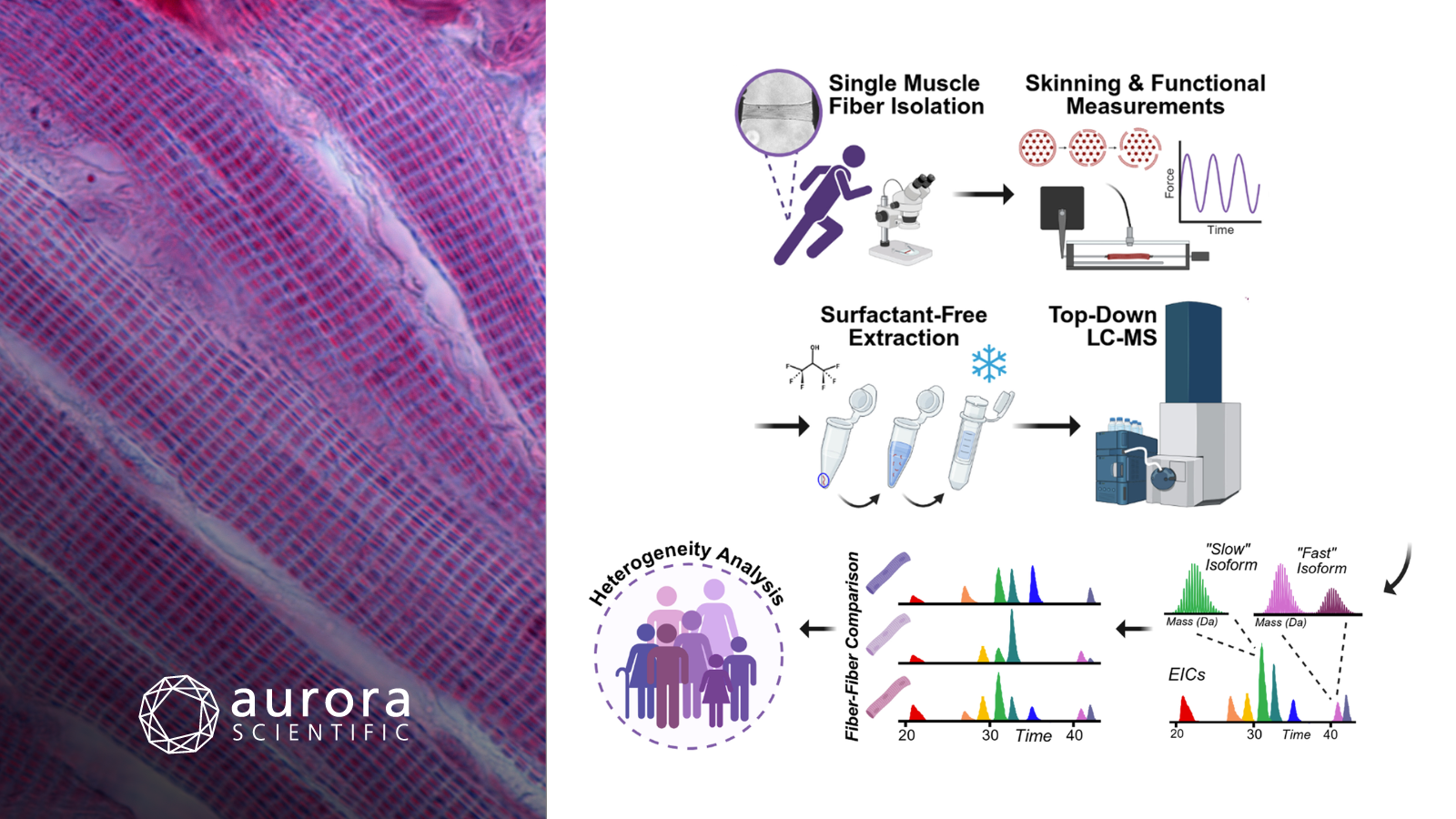
Featured image contains figures adapted from ©Wilson et al. (2026), licensed under CC BY 4.0) depicting the experimental workflow. Human single muscle fibers (hSMFs) were isolated from donor vastus lateralis muscles, chemically permeabilized, and functionally assessed. Sarcomeric proteins were then extracted using hexafluoroisopropanol (HFIP) and analyzed by top–down LC–MS for proteoform-resolved characterization, enabling unbiased assessment of intra- and inter-donor hSMF heterogeneity.
Titin’s P-Zone Domains A164–167 are Essential for Thick Filament Structural Arrangement
Titin is a key structural protein in striated muscle, playing a vital role in thick filament assembly and sarcomere function. Despite extensive research on its A-band and C-zone regions, the P-zone, which links the C-zone to the M-band, has been largely understudied. Previous structural studies have highlighted the importance of titin’s P-zone in thick filament organization, but its precise functional role remains unclear. Browne et al. (2026) addressed the gap by investigating the first four domains of titin’s P-zone (A164–A167) using a novel mouse model, revealing their critical role in thick filament structural arrangement and muscle function.
The study used genetically-engineered TtnΔA164–167 mice to examine muscle function, with successful modifications confirmed by PCR. Single EDL fibers were dissected, permeabilized, and assessed with Aurora Scientific’s 322C: High-Speed Length Controller, 403A: Force Transducer, 802D-120-TJ: Temperature Jump Permeabilized Fiber Test Apparatus, and 900B: Video Sarcomere Length system for real-time force, length and SL measurements. Force testing included passive and active protocols, cross-bridge kinetics, and calcium sensitivity, with fibers micro-knotted for stability. Single-cell experiments were then performed on an inverted microscope stage with the 1600A: Permeabilized Myocyte System.
The deletion of titin’s A164–167 domains in cardiac muscle led to a shift in titin’s epitopes, with significant changes in domain positioning, particularly in the P-zone and C-zone, towards the M-band. Thick filament length measurements revealed a slight reduction in the TtnΔA164–167 mutant (1,428 nm vs. 1,531 nm in WT), consistent with the shift in titin domains. These changes were mirrored by a shift in the positioning of cMyBP-C stripes, although their spacing remained unchanged. This study provides valuable insights into how specific titin deletions can impact sarcomere structure, highlighting the delicate balance of titin’s role in cardiac muscle function and the potential implications for disease modeling.
Top–Down Proteomics of Skinned Human Muscle Fibers Reveals Proteoform- Resolved Fiber- to- Fiber Variability
Human skeletal muscle is highly diverse, with individual fibers classified into distinct types based on contractile properties and myosin expression. However, existing methods fall short of fully capturing the molecular variability that exists within and between these fibers. Therefore Wilson et al. (2026) used top-down proteomics to analyze single human muscle fibers, revealing significant fiber-to-fiber differences in proteoform expression and abundance.
Human single muscle fibers (hSMFs) were isolated from vastus lateralis muscles and prepared for contractile measurements using Aurora Scientific’s 403A: Force Transducer and 308C: High-Speed Length Controller. After skinning, fiber proteins were extracted using a surfactant-free protocol, separated by reverse-phase chromatography, and analyzed by mass spectrometry. Data analysis involved deconvolution to quantify proteoform abundance and phosphorylation levels.
The study revealed significant fiber-to-fiber variability in isoform expression and proteoform abundance in human single muscle fibers (hSMFs) from both single and multiple donors. Notably, both intra- and inter-donor analyses demonstrated pronounced heterogeneity, with distinct differences in the composition of slow- and fast-twitch fiber isoforms and phosphorylation patterns. These findings underscore the need for single-fiber proteomics to accurately capture the molecular diversity in human skeletal muscle, enhancing the understanding of muscle biology and disease mechanisms.
Myosin-Binding Protein C Slows Cardiac Myofibril Relaxation Kinetics
Mutations in cardiac myosin-binding protein C (cMyBP-C) are a leading cause of hypertrophic cardiomyopathy (HCM), with most mutations resulting in reduced protein expression and diastolic dysfunction. However, the precise effects of cMyBP-C on myofibril relaxation kinetics remain poorly understood, particularly in relation to its cross-bridge interactions and phosphorylation-dependent regulation. Dvornikov et al. (2025) therefore used a “cut-and-paste” method to investigate the role of cMyBP-C in myofibril activation and relaxation kinetics, focusing on how its loss, phosphorylation, and mutations affect both phases of relaxation. By elucidating these mechanisms, the study provides deeper insights into how cMyBP-C modulates cardiac muscle function and how its dysfunction contributes to HCM.
Adult homozygous SpyC3 mice were treated with propranolol for five days before their hearts were excised and left ventricles harvested. Myofibrils were prepared by thawing frozen tissue and permeabilizing it with detergents, followed by digestion with tobacco etch virus protease (TEVp) to cleave cMyBP-C and produce C0-C7 fragments. Force measurements were recorded by Aurora Scientific’s 1700A: Myofibril System, in which myofibrils were attached to glass microtools, activated with calcium solutions, and precisely perfused with activating and relaxing solutions. Data were analyzed using the 600A: Real-Time Muscle Data Acquisition and Analysis System and custom Matlab scripts to fit activation and relaxation kinetics, providing insights into myofibril behavior under different conditions.
The study revealed that cleavage of cMyBP-CC0C7 from murine ventricular myofibrils accelerated both the slow and fast phases of relaxation and reduced myofilament Ca2+ sensitivity, while ligation of wild-type cMyBP-C or phosphorylation of cMyBP-C rescued these effects. Force and relaxation measurements revealed significant differences in the relaxation dynamics across different cMyBP-C manipulations. Additionally, the L348P mutation in cMyBP-C slowed both relaxation phases and increased myofilament Ca2+ sensitivity, while mavacamten treatment reduced maximal tension and accelerated relaxation, independent of cMyBP-C presence. These findings highlight the crucial role of cMyBP-C in regulating cardiac muscle relaxation and provide insight into how cMyBP-C modifications and external factors like mavacamten can influence myofibril function.
Conclusions
These studies by Browne et al. (2026), Wilson et al. (2026), and Dvornikov et al. (2025) spring forward our understanding of myofilament biology, revealing how titin structure, fiber-to-fiber proteoform variability, and cMyBP-C regulation shape muscle function and disease. Together, they highlight molecular mechanisms that keep muscle research moving into a new season of discoveries.