Our previous blog post discussed the advantages of testing muscle function in-situ or in-vivo with our 3-in-1 muscle test system. This blog post will discuss how to setup an experiment to measure muscle function in-vivo.
In-vivo muscle testing involves stimulating either the anterior crural muscles or the posterior crural muscles to elicit dorsiflexion or plantarflexion contractions respectively.
The anterior crural muscle group consists primarily of the tibialis anterior muscle (TA) and the extensor digitorum longus muscle (EDL). The posterior crural muscle group consists primarily of the gastrocnemius, soleus and plantaris muscles.
In-vivo measurements are performed by placing the anesthetized animal on a temperature regulated apparatus (mouse, and rat apparatus available). The limb (or limbs) of the animal should be shaved and sterlized.
The knee is fixed with a custom made clamp that is non-invasive to minimize damage to the animals leg and knee joint. Typically a buttressing pad or cone point set screw is used for this purpose. It is best to attempt to clamp the limb of the animal just above the knee joint itself on the fleshy portion of the hock.
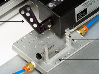
Once the leg is secured the foot can be fixed into a footplate which is attached to the shaft of a 300C series dual mode lever. The foot is attached to the footplate using adhesive tape to prevent movement within the footplate. This can be a major source of error when performing these experiments. When taping the foot, it is a good idea to first tape around the top of the foot to secure it to the pedal. Then hold the animal’s ankle with a pair of forceps and gently press the heel up against the pedal itself before applying more wrapping. This will ensure that the heel will not lift off from the pedal during the experiment.
Some iterative correction of the knee clamping and foot taping may be required before moving on to the actual experiment itself and placing of the stimulation electrodes.

Fig 1. Footplate attached to 305C motor with knee clamp on temperature regulated platform
The most difficult part of the experiment is correctly placing the electrodes so as to stimulate the desired muscles. The electrodes are connected to our 701C bi-phase stimulator. The stimulator is controlled by our Dynamic Control and Analysis software which also acquires the torque measurements from the 300C footplate.
Electrode placement:
- For stimulation of anterior compartment muscles, including the tibialis anterior and the extensor digitorum longus muscle, place the electrodes directly over (surface electrodes) or subcutaneously either side (needle electrodes) of the animal’s deep fibular nerve, which is a distal branch off of the common peroneal nerve. Alternatively the common peroneal nerve can also be stimulated.
- For stimulation of the posterior compartment muscles, including the gastrocnemius, soleus and plantaris muscles place the electrodes directly over (surface electrodes) or subcutaneously (needle electrodes) either side of the animal’s sciatic nerve. The tibial nerve can also be stimulated. In some circumstances and where possible (terminal experiments), it may also be useful to sever the common peroneal nerve to prevent recruitment of the anterior muscles
- Optimisation of the position of the electrodes should be ascertained for every experiment to ensure that the desired muscles are stimulated and the maximum isometric twitch torque is achieved. Once the optimal position of the electrodes is ascertained the electrodes should be securely kept in place.
As with isolated and in-situ muscle experiments, the stimulation voltage/current should be optimized to obtain peak isometric torque before an experiment is performed. Unlike ex-vivo and in-situ experiments the muscle and tendon is still completely attached to the limb of the animal and therefore optimal resting length does not have to be ascertained for each experiment. However, optimal position of the leg with respect to the angle of the knee/leg should be determined. A 90 degree angle is an appropriate place to start.
The experimental protocol can now be performed. The animal should be regularly checked for response to stimulation in order to ensure the anesthetic level is sufficient. The anesthesia should be adjusted as needed. Once the protocol is finished the animal can be removed from anesthesia and allowed to recover.