September, for many students around the world, means a return to the classroom and the laboratory. For us at Aurora Scientific, September always means a visit to the European Muscle Conference hosted by the European Society for Muscle Research. This is an international meeting showcasing some of the finest work on myofilament structure and function along with muscle energetics, disease, and, of course, translational work. Understanding the mechanisms of muscle contraction at the level of the myofilament and myofibrillar proteins requires the use of a vast array of experimental techniques, with our contractility equipment playing a small part. Other techniques such as AFM, histology, protein gels, and X-ray diffraction are indispensable. Here, we present you with some recent publications which make use of these techniques, particularly X-ray diffraction, to further our understanding of the myofilament.
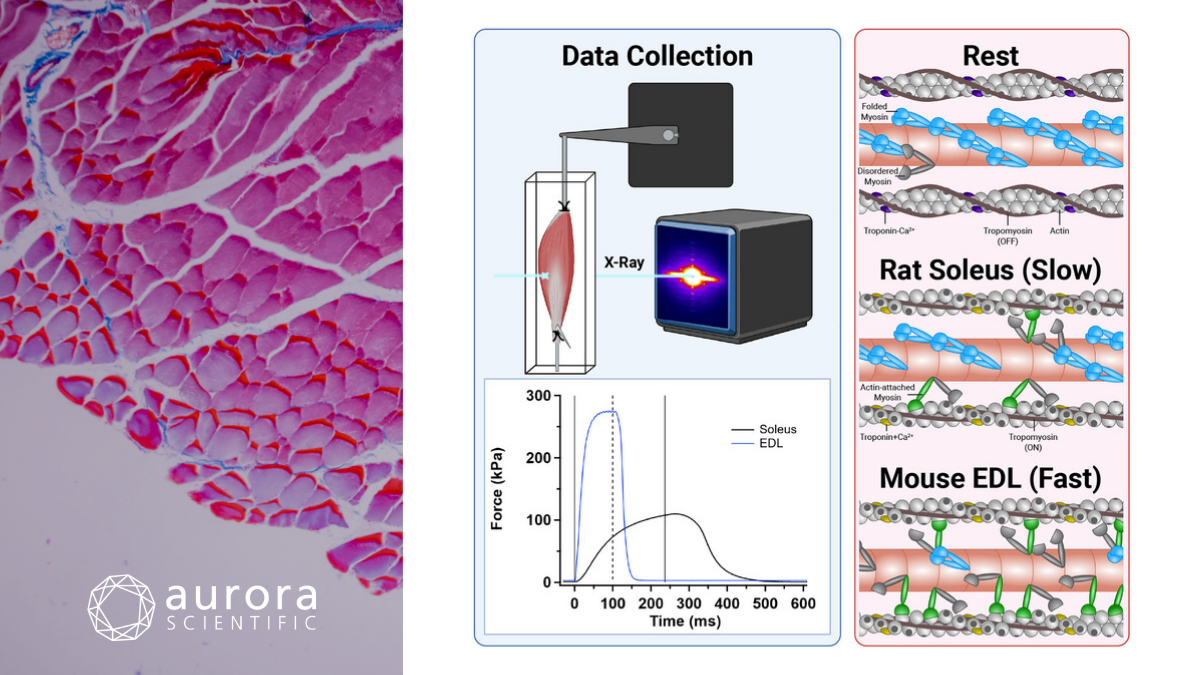
Featured image contains figures adapted from Hill et al. (2026), licensed under CC BY 4.0, depicting the experimental setup and structural differences between slow and fast skeletal muscle contraction. Time-resolved small-angle X-ray diffraction (SAXD) patterns were used to study rat soleus muscle during fixed-end twitch and tetanic contractions. In resting muscles, most myosin motors (blue) are folded against their tails in a helical array on the surface of the thick filaments (pink). At the plateau of a fixed-end tetanus, rat soleus slow muscle generates lower force than mouse extensor digitorum longus (EDL) fast muscle, with fewer myosin motors attached to thin filaments (grey) in the perpendicular force-generating conformation (green), and more motors remaining in the folded OFF state (blue).
Distinct distributions of myosin motor conformations during contraction of slow and fast skeletal muscle
In nature, slow-twitch muscle notably has been shown to produce movement with higher metabolic efficiency than fast-twitch muscles. When comparing fast and slow-twitch muscles in the same species, the ADP consumption cost can be 5 times higher in fast-twitch muscle compared to the slow. Historically, mechanical calculations implied that slow muscle required more attached cross-bridges to maintain isometric tension than fast-twitch muscle, which is entirely paradoxical to the historical energetics assessments in the literature. In this paper, Hill et al. (2026) used high-precision small-angle X-ray diffraction (SAXD) to precisely determine how many myosin motors remain attached to the actin filament during the period of active contraction. Could the findings resolve this biophysical contraction?
In the experiment, the rat soleus was surgically removed in the usual fashion and prepared for standard in-vitro contractile mechanics work. A 300C-LR: Dual Mode Muscle Lever was used to record contractile force, and a 701C: Electrical Stimulator was used to electrically activate the muscle to perform a contraction train protocol. In lieu of a traditional bath like our 800C: in-vitro muscle apparatus, the authors used a specially designed bath with mylar windows closely adjusted to the width of the muscle itself. The bath/trough was sealed so the solution did not leak and the bath was suspended vertically in the path of the ID02 beamline at the ESRF in Grenoble and at the I22 beamline at DLS. The chosen beamlines were meant to maximize the spatial resolution when assessing the diffraction pattern. Exposure time and the distance from sample to detector varied for twitch and tetanus experiments.
Contrary to historical mechanical calculations that implied that slow muscle required more attached cross-bridges to maintain isometric tension, Hill et al. (2026) discovered the exact opposite using high-precision small-angle X-ray diffraction. This structural distribution perfectly explains how slow muscle maintains long-term postural tension at a low metabolic cost. During an active isometric contraction, slow-twitch muscles have significantly fewer myosin motors attached to actin than fast-twitch muscles because a much larger pool of motors remains safely sequestered in an “OFF” state on the thick filament backbone. Because the majority of motors in slow muscle stay tucked away in the energy-saving folded conformation, ATP consumption remains incredibly low. The early structural changes in slow muscle occur at roughly the same amplitude in a brief twitch as they do in a prolonged tetanus. This indicates that slow muscle activation relies on a highly responsive, direct pathway triggered by thin-filament activation rather than the heavy mechanical stress required to “wake up” fast-twitch thick filaments.
Kbtbd13 knockdown restores muscle function in a clinically relevant mouse model of nemaline myopathy type 6
Nemaline myopathy type 6 (NEM6) is a rare congenital muscle disorder characterized by muscle weakness and a peculiar “slowness” of muscle movement caused by impaired relaxation kinetics. Research indicates that mutations in the KBTBD13 gene cause the protein to bind abnormally to actin, stiffening the muscle’s thin filaments and rendering muscles unable to relax quickly. Researchers used a Kbtbd13:p.Arg408Cys knockin mouse model in Galli et al. (2026), which closely matched the phenotype of human NEM6 patients. To understand the mechanism of the disease, researchers used low-angle X-ray diffraction in conjunction with functional contractility assays. Since it has been previously shown that knocking out the KBTBD13 protein does not cause adverse effects on intact muscle mechanics, uncovering the mechanism of NEM6 myopathy could lead to a real, viable therapeutic pathway.
Whole mouse muscle in-vitro mechanical measurements were done on both the knockin mice and wild-type animals using an Aurora Scientific 1200A: Isolated Muscle Test system. It was shown that the phenotype of Soleus muscle was more affected than EDL muscle, and that it occurred at earlier time points in the disease progression. To better understand the structure, the researchers used small-angle X-ray diffraction at beamline 18D at BioCAT in Argonne, USA, from both EDL and Soleus in 9-month-old mice (the age at which the severity of the phenotype appeared to peak). The muscles were electrically stimulated and diffraction patterns were collected during relaxation and maximal activation. These X-ray measurements showed a reduction in actin spacing from relaxed to activated states, and an increase in stiffness in Soleus muscle.
The mutated KBTBD13 protein binds directly to actin, and thus these structural changes of the thin filament lock the actin-tropomyosin complex into a state that slows and perturbs the normal kinetics of myosin cross-bridge detachment. To further test this, the researchers evaluated the mice treated with the AAV9-shRNA gene therapy, which was used to knock down or silence the KBTDB13 protein. Actin spacing and stiffness were largely restored. Would fixing the molecular structure of the thin filament fully restore physical muscle function? Across a wide variety of in-vitro and in-vivo functional assays, including contractility, treadmill running and morphological assays, outcomes were dramatically improved by a single viral vector injection, offering great promise for a potential human therapeutic pathway.
Conclusions
These studies by Hill et al. (2026) and Galli et al. (2026) demonstrate how the small-angle X-ray diffraction technique can contribute to our basic understanding of the biophysical properties of the myofilament, but also have the ability to validate the potential of translational therapeutics. We are proud to continue to support researchers who are adding the small-angle X-ray technique to their experimental toolbox, and we have a growing line of chambers and equipment that can be used with this technique, including the new 840A: BioVault. We are proud to support this amazing research and look forward to expanding our line of instruments specifically designed for this technique.
If you have any suggestions for future publication reviews, please reach out to us directly with your suggestions, and we would be glad to feature your own research or that of a colleague’s!