The kinematic activity of skeletal muscle helps inform the physiology behind muscle contraction and force production. The equipment needed to induce force, mount samples, and capture structural changes are among Aurora Scientific’s line of instrumentation and is highlighted in some recent publications in this review.
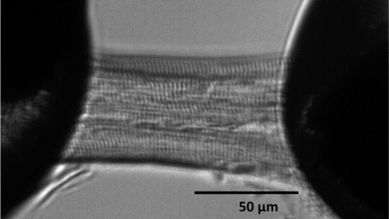
Featured image (©Greenman et al. 2022, licensed under CC BY) showcases a single cardiomyocyte mounted using Aurora Scientific equipment with our 406A Force-Transducer attached to the left pin.
Ultrastructural and Kinetic Evidence Support That Thick Filaments Slide Through The Z-Disc
Muscle activation and force generation involve the overlap of thick and thin filaments in a sliding motion that exists in a force-length relationship (FLR). How the filaments work at such small scales is not clear though it commonly believed that thick filaments are compressed at the Z-disc. Tomalka et al. (2022) sought to determine if this is indeed the case, and to give some evidence in support of the theory.
The authors were able to provide evidence to confirm hypotheses about thick filament sliding through the Z-disc. They reported ultrastructural, kinetic, and kinematic evidence through their exploration. They prepared muscle from Wistar rats and completed their kinetic experiments using our 1400A Permeabilized Fiber System, Sarcomere length was measured and used to set optimal length of fibers using our 901B High-Speed Video Sarcomere Length system. The authors concluded that their results were not compatible with the theory of compression of the thick filament but did support thick filament sliding through the Z-disc. Certainly, these findings will inform our understanding of skeletal muscle physiology.
Treadmill Running Increases The Calcium Sensitivity Of Myofilaments In Diabetic Rats
The benefits of exercise cannot be overstated. However, in type 2 diabetes the response to exercise is poor because of reduced cardiac performance causing exercise intolerance. This is attributed to attenuated cardiomyocyte contractility. The diabetic heart must maintain cardiac output by elevating resting heart rate. Cardiac output struggles to meet increased oxygen demand because of impaired stroke volume. Greenman et al. (2022) sought to determine if myofilament calcium sensitivity can be altered with exercise training since it is known to improve cardiac reserve in diabetes.
The authors used diabetic and non-diabetic rats in an 8-week treadmill running training protocol. Cardiomyocytes were examined using our 406A Force Transducer and sarcomere length was captured using our 900B High-Speed Video Sarcomere Length system. The findings of the study were that training increased myofilament calcium sensitivity but training did not alter phosphorylation of myofilament proteins. While this is an unconfirmed possible mechanism that can compensate the reduction in cardiac reserve in diabetes, the study suggests that regular exercise may improve diabetic heart performance by targeting myofilament contractility.
Titin Force In Muscle Cells Alters Lattice Order, Thick And Thin Filament Protein Formation
Length-dependent activation (LDA) summarizes the process through which muscles can exhibit a range of contraction tuning based on demand. The sarcomeric protein titin maintains connections within the sarcomere that are believed to drive LDA. However, the control scheme for LDA is not fully understood. Hessel et al. (2022) sought to track mechanical and structural changes both before and after a reduction in titin-based force.
The authors used a mouse model and X-ray diffraction to cleave titin protein. They prepared the fibres using our 402A Force Transducer, and 322C-I High-Speed Length Controller. The force and length data were collected using our 600A Real-Time Muscle Data Acquisition and Analysis System. The authors found that when titin was cleaved by 50%, LDA was attenuated in thick filaments. It appears titin plays a regulatory role in LDA structural features. The authors further demonstrated that thin filaments also contained length-dependent priming through protein binding between thick and thin filaments in resting muscle. Their results will be important in future studies of LDA especially where LDA impairment may be a critical factor in muscle disease.


