Press lightly on the back of your hand and you feel touch. Press harder, or with something sharp, and at some point, that sensation turns to pain. Where does that line sit, and which nerve fibres draw it? This publication review gets straight to the point with two studies that tackle the question the same way: by pressing the skin with precisely controlled force and watching which sensory neurons respond, and how strongly. One maps the answer across a dozen types of sensory neurons in mice; the other zooms in on single nerve fibres in humans.
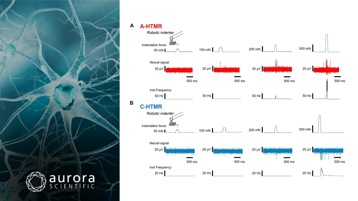
Featured image contains a figure adapted from Bouchatta et al. (2026), licensed under CC BY 4.0, depicting example responses of A) an A-fibre high-threshold mechanoreceptor (A-HTMR) and B) a C-fibre high-threshold mechanoreceptor (C-HTMR) to skin indentation delivered by a robotic indenter. Traces show indentation force markers, the raw neural signal and instantaneous firing frequency in response to 60, 100, 260 and 1000 mN forces.
A mouse DRG genetic toolkit reveals morphological and physiological diversity of somatosensory neuron subtypes
Sensory neurons in the dorsal root ganglia (DRG) detect virtually all types of stimuli from a light breeze to a painful pinch. Mapping how their many subtypes share that job has been difficult, partly because there were no tools to target many of them individually, particularly the CGRP-expressing neurons often grouped together as “peptidergic nociceptors.” To fill this gap in research, Qi et al. (2024) sequenced roughly 40,000 mouse DRG neurons, identified at least 15 transcriptionally distinct subtypes, and generated new mouse lines to label them. With genetic access in hand, could they pin down how each subtype responds to force?
The team performed in vivo calcium imaging of DRG neurons while stimulating hairy skin on the mouse thigh. Aurora Scientific’s 300C-I: Dual-Mode Indenter, fitted with a custom 200-µm tip, delivered 0.5-second step indentations from 1 to 75 mN to each neuron’s receptive field, and longer, 3-second steps revealed how each subtype adapted to sustained pressure. Because every subtype received the same calibrated stimuli, their force thresholds could be compared directly. Thermal responses were also tested on the same patch of skin using a custom Peltier device.
The result was a clear division of labour. Most low-threshold mechanoreceptors (LTMRs) responded to just 1–5 mN, with Aβ RA-LTMRs mostly under 10 mN, and their responses levelled off between 20 and 40 mN. Six high-threshold subtypes progressively switched on later: MRGPRD+ and MRGPRB4+ neurons mostly at 5–20 mN, BMPR1B+ neurons around 20 mN, CYSLTR2+ and MRGPRA3+ neurons at 20–40 mN, and SMR2+ neurons mostly above 40 mN. All six kept firing throughout sustained indentation, while two thermally tuned populations, TRPM8+ and SSTR2+ neurons, ignored indentation altogether. Together, these thresholds tile the full range of forces tested, supporting a population code in which more subtypes join in as pressure rises.
The SMR2+ neurons stood out. These lightly myelinated, CGRP+ neurons responded only to noxious force, and their endings reach into the epidermis, challenging the common assumption that CGRP+ fibres there are C-fibres. The findings also led the authors to question the catch-all label “nociceptor,” since most high-threshold subtypes also respond to innocuous forces, and to propose naming subtypes by their physiology instead.
Coding of mechanical pain by myelinated and unmyelinated nociceptors in human hairy skin
Textbook models split the work: fast, thickly myelinated Aβ fibres carry touch, while pain travels more slowly along thinly myelinated Aδ and unmyelinated C fibres. Evidence from animals and humans has been chipping away at that divide. The same research group previously identified “ultrafast” A-fibre high-threshold mechanoreceptors (A-HTMRs) in human skin, which conduct at touch-like speeds and evoke pain when activated. Building on that discovery, Bouchatta et al. (2026) set out to measure how these fibres encode force, and how they compare with their slower C-fibre counterparts (C-HTMRs).
Using microneurography, the researchers recorded single nerve fibres from the radial nerve of awake, healthy volunteers. To assess force coding, they used Aurora Scientific’s 305C-I: Dual-Mode Indenter as a robotic indenter, fitted with a 1-mm flat tip and mounted on an X-Y plotter to make it easier to position during recordings. It delivered 0.2-second indentations at five forces, from 20 to 1000 mN, to each fibre’s receptive field. In separate psychophysical testing, 21 participants rated the unpleasantness of the same five forces, again delivered by the Aurora indenter.
Both A- and C-HTMRs fired more as force increased, in a linear stimulus–response relationship. However, A-HTMRs responded significantly more vigorously on every measure: spike count, mean firing frequency and peak frequency. They also conducted at about 48 m/s, on par with touch fibres, so mechanical pain can be signalled as quickly as touch. Additionally, while field-type touch fibres plateau once forces turn painful, as the group showed in earlier work, HTMRs kept scaling.
The perceptual results matched. Unpleasantness rose with force, and “sharp” was the most common descriptor, followed by “stabbing.” Weak electrical stimulation of a single A-HTMR through the recording electrode produced a well-localized, sharp, stinging pain right where its receptive field lay. The authors also credit part of the advance to the stimulator itself: unlike the hand-applied von-Frey monofilaments used in earlier studies, the robotic indenter’s force was verified by a built-in sensor and delivered through the same tip at every force. Their figures even describe the resulting sensations as “Aurora-induced percepts.”
Conclusions
These studies by Qi et al. (2024) and Bouchatta et al. (2026) get right to the point. In mice and in humans, touch receptors max out early, while high-threshold neurons keep scaling into the painful range, and myelinated high-threshold fibres emerge as specialists for intense force. Getting there took the same thing in both labs: precisely controlled, repeatable force, from 1 mN on a mouse’s thigh to a full newton on the back of a human hand. The instruments used here have since been succeeded by our 300E-I: Dual-Mode Indenters, and we are proud to support this research. Touch taps out early; nociceptors keep counting.
If you have any suggestions for future publication reviews, please reach out to us directly with your suggestions, and we would be glad to feature your own research or that of a colleague’s!