As we launch full force into 2024, studies exploring the molecular underpinnings of force production in mice have blazed the trail of functional muscular research. From calcium dynamics during muscle contraction, to muscle impairments in myotonic dystrophy, and the effects of running on muscle mass in a dystrophic mouse model, January’s novel insights into muscle function and regulation have important implications on our understanding of muscle physiology and health.
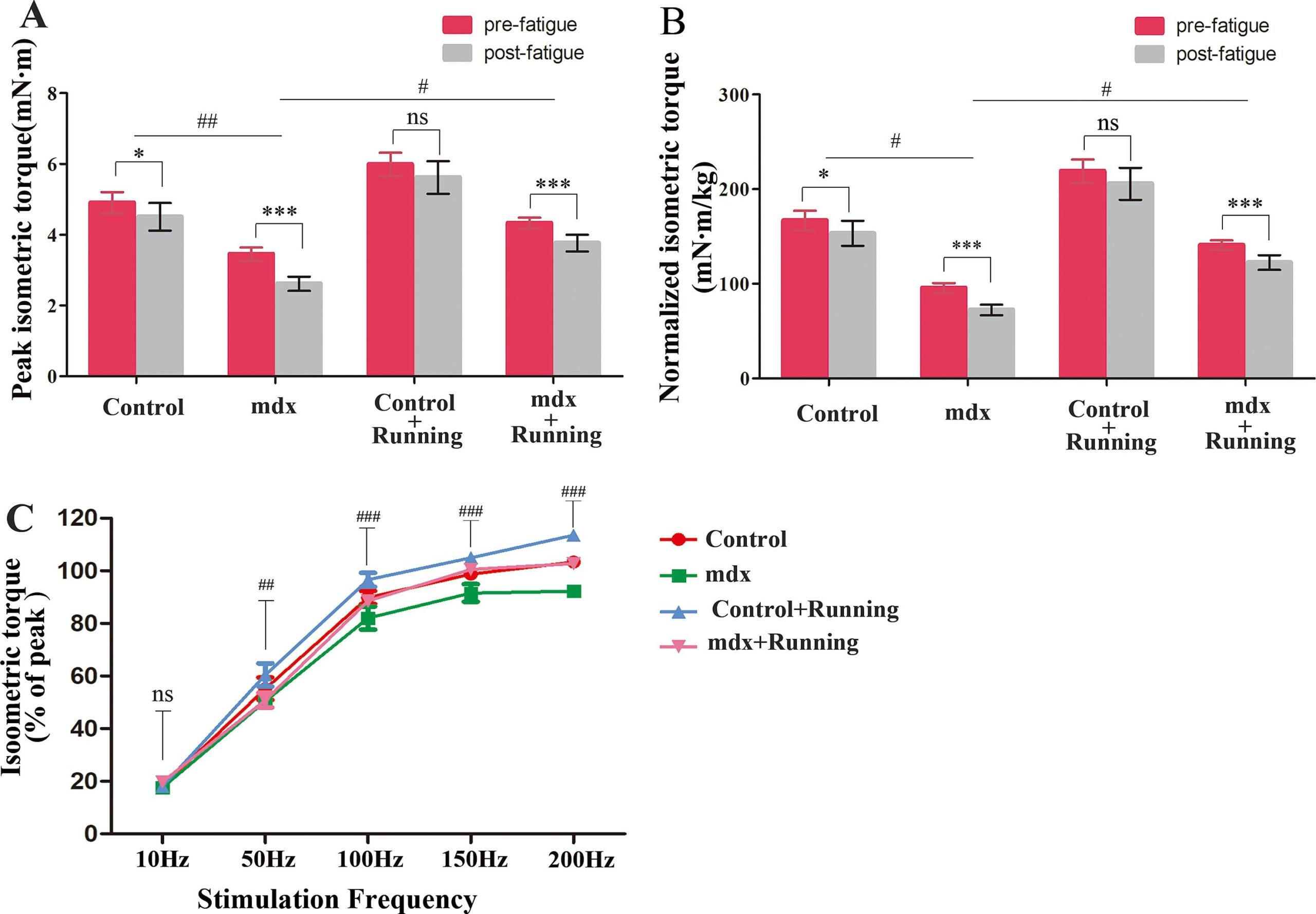
Featured image (Zhou et al. (2024), ©2024 Elsevier B.V. All rights reserved.), demonstrating how running can promote fatigue resistance in mouse skeletal muscle. A,B) peak isometric torque pre- and post-fatigue. C) Relative peak isometric torque-frequency curve post-fatigue.
Ca2+ and force during dynamic contractions in mouse intact skeletal muscle fibers
Calcium’s crucial role in muscle activity has been cemented by the well-established dependence of muscle contraction on cytosolic calcium. On a molecular level, sarcolemmal depolarization triggers the release of calcium ions, which bind to troponin and effectively expose actin sites. This allows for myosin heads to bind to actin and form actomyosin cross-bridges, thereby resulting in muscle contraction. In this way, when the frequency of sarcolemmal depolarization increases, cytosolic calcium concentrations also increase, allowing muscles cells to contract with increasing force.
Although several studies have consistently reported that 100 Hz is required to induce maximal force in isometric contractions, the force-frequency relationship in dynamic contractions remains critically understudied. As such, Fukutani et al. (2024) sought to investigate the interplay between force and cytoplasmic free calcium concentration ([Ca2+]i) in intact mouse single flexor digitorum brevis (FDB) fibers. To achieve this, the fiber was mounted on Aurora Scientific’s 801C Small Intact Muscle Apparatus, which was placed on an inverted microscope. Together with the 403A Force Transducer and 315C High-Speed Length Controller, this muscle system enabled force measurements exerted by the fibers during isometric and dynamic contractions.
Interestingly, while tetanic [Ca2+]i remained similar across contraction types, force differences were notably observed. Specifically, the force attained at the 30 Hz stimulation relative to the 100 Hz stimulation was smaller in the concentric than in the isometric contraction. Taken together, these findings demonstrate that myofibrillar calcium sensitivity, rather than changes in [Ca2+]I, contributes to force variations during dynamic muscle contractions.
Verapamil mitigates chloride and calcium bi-channelopathy in a myotonic dystrophy mouse model
Myotonic dystrophy type 1 (DM1) is an autosomal dominant genetic disorder characterized by myotonia and progressive muscle weakness. On a genetic level, DM1 is caused by the expansion of an unstable trinucleotide repeat of cytosine-thymine-guanine (CTG) in the DMPK gene, which leads to the accumulation of toxic RNA. This toxic RNA effectively disrupts cellular function, leading to misregulated alternative splicing and disrupted RNA processing.
While individual splicing defects have been studied to decode the DM1 phenotype, it is largely unknown what underlies the progressive muscle weakness shared across patients. As such, Cisco et al. (2024) sought to delineate the muscle phenotype by expertly employing exon or nucleotide deletion of genes related to muscle excitation-contraction coupling in mice. Using Aurora Scientific’s 1200A system, they went on to perform ex vivo EDL muscle contraction measurements to assess functional force deficits. Upon testing, they found that mimicking chloride and calcium bi-channelopathies resulted in significantly reduced lifespans, myotonia, mobility and respiratory impairment in their mice. Interestingly, after administering a calcium channel blocker, called Verapamil, and reassessing the mice, they noted significantly improved survival, force generation, myotonia, and respiratory function. These findings suggest that Ca2+/Cl– bi-channelopathy contributes to muscle impairment in DM1, and that these muscle impairments could potentially be ameliorated by clinically available calcium channel blockers.
Running improves muscle mass by activating autophagic flux and inhibiting ubiquitination degradation in mdx mice
Duchenne muscular dystrophy (DMD) is a severe, progressive disorder caused by mutations in the dystrophin gene, located on the X chromosome. These mutations lead to the absence or dysfunction of a functional dystrophin protein, ultimately leading to muscle degeneration and weakness.
Within the field, the mdx mouse has been widely employed to study the molecular mechanisms underlying DMD; however, varying effects of exercise on muscle function have been reported throughout the years. To address this, Zhou et al. (2024) investigated the effects of 10 weeks of treadmill training on skeletal muscle in control and mdx mice. They used Aurora Scientific’s 1300A Whole Animal System to measure maximal isometric torque in mice, pre- and post-administration of their fatigue protocol. Interestingly, their results revealed that running exercise significantly improved muscle mass, strength, endurance, and anti-fatigue ability in mdx mice, effectively reversing the pathological state of skeletal muscle damage, and promoting regeneration. At the molecular level, running inhibited the ubiquitination and degradation of muscle protein, suppressed AKT activation, decreased phosphorylated FoxO1 and FoxO3a, and restored autophagic flux. Furthermore, they observed comprehensive promotion of fast and slow muscle fibers, suggesting that running functionally enhances muscle strength and endurance. Collectively, these findings indicate that progressive, low-medium-speed, and low-frequency running can inhibit muscle protein degradation, promote protein reuse and accumulation, and improve skeletal muscle mass in mdx mice.
Conclusions
Recent advances in muscle physiology have uncovered the molecular elements at play in muscle force production. These studies by Fukutani et al. (2024), Cisco et al. (2024), and Zhou et al. (2024) have performed a molecular deep dive into the role of calcium, chloride and calcium bi-channelopathies, and exercise in different contexts of muscle health and disease. Together, their findings contribute to our understanding of muscle physiology and the promising functional improvements that can arise from interventions in muscle impairments.


