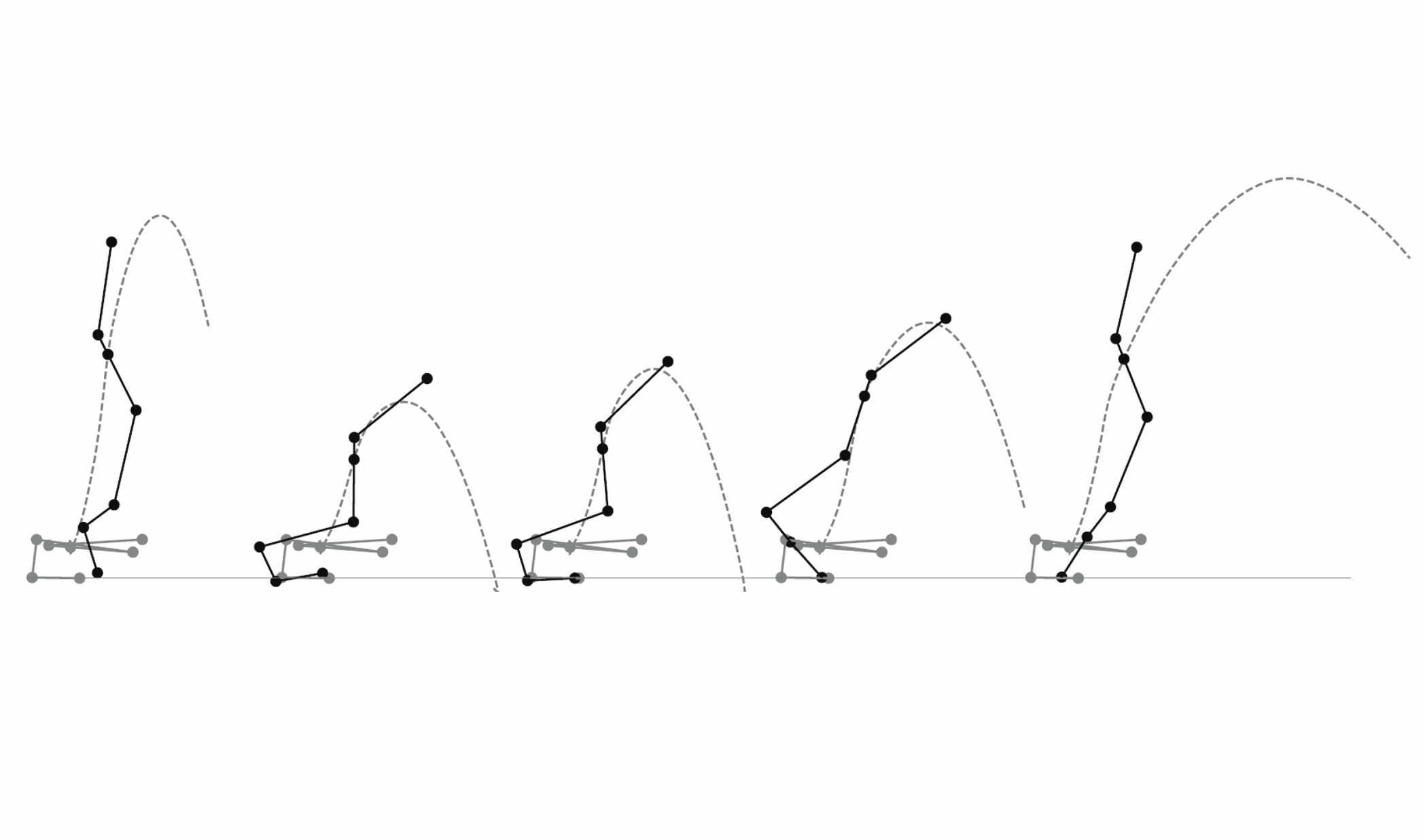
Experimentation in vitro enables physiologists to control neuromuscular stimulation and strain patterns to help determine how the nervous system modulates musculoskeletal physiology. Recently, computerized simulations have been advanced to be able to test the effects of musculoskeletal parameters, such as elasticity and inertia, on in vitro muscle samples. Furthering these techniques and current advances in muscle-hardware feedback, hybrids of in vitro physiology and musculoskeletal simulation have been developed. These advancements in physiologically-relevant simulations provide a better understanding of how evolutionary changes in limb architecture influence the mechanical demands of muscle. In addition, these simulations allow for broader applications in muscle physiology research, such as testing tissue samples to better isolate and understand the effects of injury or disease on locomotion.
Featured Image sourced from https://doi.org/10.1242/jeb.210054In vitro virtual reality: an anatomically explicit musculoskeletal simulation powered by in vitro muscle using closed-loop tissue–software interactionTo demonstrate the utility of virtual simulations for muscle physiology research, Richards & Eberhard (2020) tested the effects of muscle stimulation on altering limb anatomy by modifying a muscle’s anatomical origin. In this publication, they describe the methods, limitations, and data that demonstrate the utility of ‘in vitro virtual reality’ (in vitro-VR) for capturing the effects of virtual skeletal manipulation on the force-length dynamics of a muscle sample. The in vitro-VR used here is a hybrid of two components; 1) a 2D musculoskeletal simulation model that computes joint kinematics in response to joint torque inputs and, 2) an in vitro muscle–tendon tissue sample that is mounted to a custom ergometer instrumented with our 305C-LR muscle lever system. This lever system measures force at different lengths of the muscle-tendon sample during contraction. This measured force is converted and fed back to the 2D simulation model in the closed loop to update the model. In vitro-VR can reproduce more realistic in vivo conditions to help validate recent muscle models that account for traditionally neglected features such as tissue mass and 3D anatomical structure.A novel ex vivo protocol to mimic human walking gait: implications for Duchenne muscular dystrophyBukovec, et al. (2020) used a physiologically-relevant simulation to develop a novel ex vivo mouse model to mimic human walking gait. In this publication, they discuss the implications of this research for Duchenne muscular dystrophy (DMD). To better understand eccentric muscle contractions during gait, and ultimately to better understand their effects in dystrophic muscle, the mdx mouse (a DMD model that lacks dystrophin) is used here in a scaled-down simulation of human soleus muscle during gait. This simulation protocol is used to explore the possibility that DMD muscle is exceptionally susceptible to eccentric stresses and strains. Aurora Scientific DMC software (615A), dual-mode lever system (300C-LR) and electrical stimulator (701C) were used to finely control muscle length changes to produce a force pattern that correctly mimicked the gait cycle from the human simulations. In this study, soleus muscles from wild-type and mdx mice were subjected to an ex vivo gait protocol. This protocol was well-tolerated by both groups and could assess simulated human movement in a mouse muscle. This novel assessment tool can be used to examine the effects of eccentric contractions on DMD muscle to further the advancement of Duchenne muscular dystrophy research.


