June arrives with the energy of long days and summer warmth, a season made for movement and momentum. In the same spirit, exercise science is flexing its intellectual muscle, uncovering how physical activity shapes muscles, brains, and overall physiology. Off the heels of Aurora Scientific’s attendance at the 20th International Biochemistry of Exercise Conference (IBEC), we’re excited to spotlight the latest in exercise research. From volumetric muscle loss and dystrophic adaptations to exercise-driven cognitive benefits, this month’s publication review highlights how voluntary, forced, and swimming exercise keep both tissue and knowledge in peak condition.
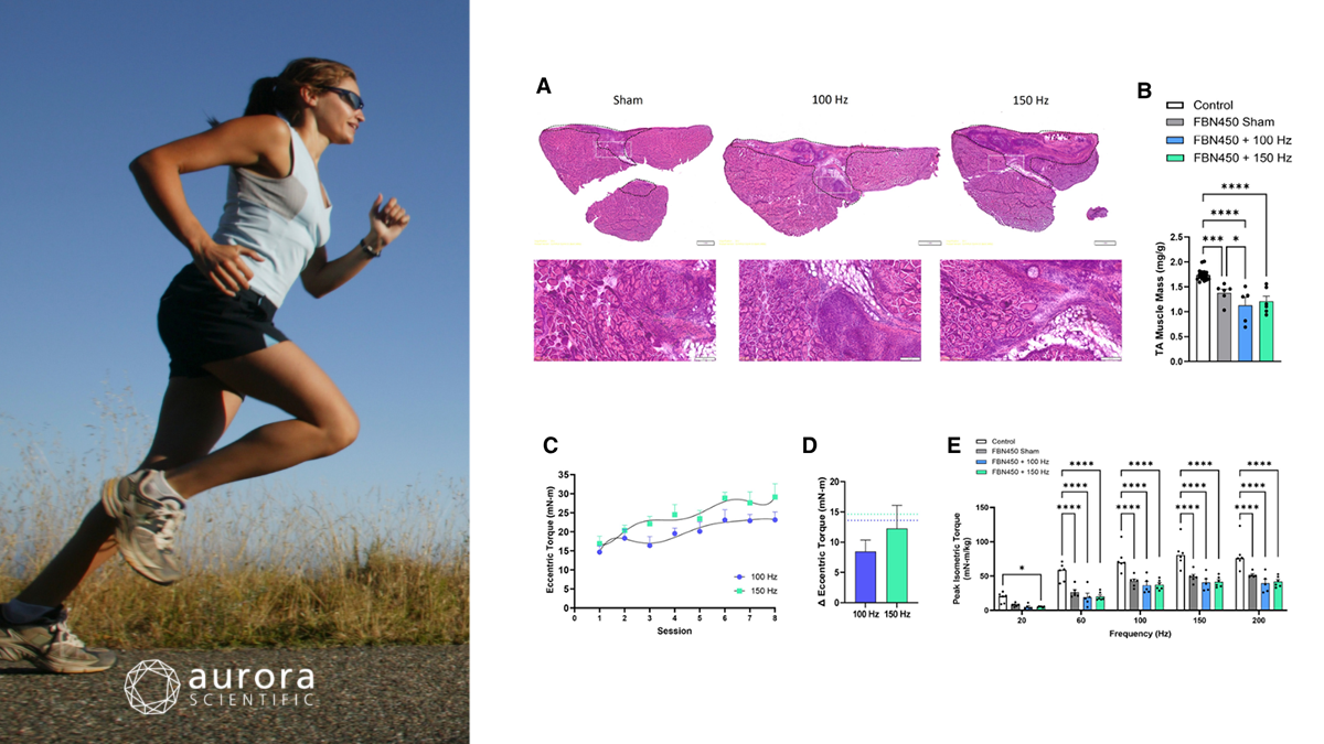
Featured image (photo of white rat on green surface from Alma Grigorita via Canva.com with figures adapted from ©Ziemkiewicz et al. (2026), licensed under CC BY 4.0) depicting A) H&E-stained muscle sections 42 days post-injury with dashed yellow lines indicating the defect region and white boxes showing magnified areas. B) VML-injured TA muscle mass measured 42 days post-injury (n = 5–6/group). C) Eccentric torque increased with 150 Hz stimulation and across bouts. D) Change in eccentric torque between bouts 1 and 8 with no differences between 100 Hz and 150 Hz programs with hydrogel treatment; dotted lines indicate values from untreated VML-injured muscles reported by Ziemkiewicz et al. (2023). E) Peak isometric torque measured and normalized to body weight. Statistical analyses were performed using two-way ANOVA or unpaired t-tests, with significance indicated by * p < 0.05, *** p < 0.001, and **** p < 0.0001.
Application of Fibrin-Laminin Hydrogel Concurrent with Electrically Stimulated Eccentric Training Hinders Recovery in Volumetric Muscle Loss
Volumetric muscle loss (VML) presents a significant clinical challenge, as the excision of muscle tissue and its supporting elements leads to chronic functional deficits that are poorly addressed by current therapies. Previous work has demonstrated that fibrin-laminin hydrogels (FBN450) can enhance muscle regeneration, while electrically stimulated eccentric training (EST) promotes hypertrophy and functional recovery in injured muscles. However, whether the combination of these regenerative and rehabilitative strategies produces additive or synergistic benefits remains unclear. Therefore, Ziemkiewicz et al. (2026) investigated the concurrent application of FBN450 and EST in a rodent VML model to determine if this combinatorial approach could maximize structural and functional muscle repair.
Adult male Lewis rats with tibialis anterior VML injuries were treated with fibrin-laminin (FBN450) hydrogels, followed two weeks later by electrically stimulated eccentric training (EST) at 100 or 150 Hz. Muscle function was assessed using Aurora Scientific’s 1305A: 3-in-1 Whole Animal System for Rats, allowing precise peroneal nerve stimulation and measurement of isometric and eccentric forces. Histology, fibrosis staining, and RT-qPCR were performed to evaluate fiber structure, tissue remodelling, and gene expression.
FBN450-treated VML muscles showed persistent deficits in mass and myofiber size, with increased cellular infiltration at the defect site. Lower peak isometric and eccentric torque were observed in hydrogel-treated muscles, with EST failing to restore function to prior levels. Fiber analysis showed reduced type 2B fiber size and smaller 2A/2X fibers, while gene expression indicated heightened inflammatory signalling and collagen 1, but no increase in myogenic markers. Collectively, these results suggest that combining FBN450 hydrogels with EST does not enhance muscle regeneration or functional recovery, highlighting the need for alternative strategies to improve outcomes following volumetric muscle loss.
Exercise alleviates cognitive dysfunction in Alzheimer’s disease mice via skeletal muscle-derived extracellular vesicles that enhance plaque clearance by microglia
Alzheimer’s disease (AD) is a progressive neurodegenerative disorder characterized by cognitive decline, yet effective strategies to slow or reverse its progression remain limited. Physical exercise has been shown to improve cognitive function and modulate brain health, in part through the release of muscle-derived factors, but the mechanisms remain incompletely understood. Emerging evidence suggests that skeletal muscle-derived extracellular vesicles (SKM-EVs) may serve as endocrine mediators, yet their role in AD and microglial activation has not been investigated. To address this, Lin et al. (2026) examined whether exercise-induced SKM-EVs enhance microglial clearance of amyloid-beta and improve cognitive function in AD mice, while identifying key molecular cargo responsible for these effects.
APP/PS1 (AD mice) and wild-type mice underwent a 4-week swimming regimen, with or without PLX5622 treatment, followed by behavioural, histological, and molecular analyses to assess cognitive function, microglial activity, and amyloid-beta clearance. Skeletal muscle adaptations were evaluated ex-vivo using Aurora Scientific’s 1200A: Isolated Muscle System for Rodents, which measured contractile properties, force-frequency responses, and fatigue. Conditioned media from these muscles were then collected to isolate extracellular vesicles for downstream analyses.
Swimming exercise in APP/PS1 mice reduced amyloid plaque burden, improved cognitive performance, and enhanced DAM (disease-associated microglia) activation, as shown by increased phagocytosis, microglial polarization, and expression of Trem2 and p-SYK. Depletion of microglia with PLX5622 abolished these exercise-induced cognitive benefits, confirming that microglial activation is necessary for the effects. Exercise was shown to increase skeletal muscle EV (extracellular vesicle) secretion, which could cross the blood–brain barrier and activate microglia, with miR-378a-3p identified as the key effector miRNA mediating these effects. Overall, these findings reveal that exercise improves cognitive function in AD mice via a skeletal muscle–microglia axis, highlighting EV-mediated inter-organ communication as a potential therapeutic mechanism.
Volitional exercise elicits physiological and molecular improvements in the severe D2.mdx mouse model of Duchenne muscular dystrophy
Duchenne muscular dystrophy (DMD) causes progressive muscle wasting and weakness, and current therapies are limited by mutation specificity and side effects. While moderate exercise benefits mild C57.mdx mice, its effects in the more severe D2.mdx model remain unclear. Mattina et al. (2026) investigated whether voluntary wheel running (VWR) could improve muscle and mitochondrial health in D2.mdx mice without worsening dystrophic pathology.
Male D2.mdx and wild-type mice underwent 8–10 weeks of voluntary wheel running or remained sedentary, followed by body composition, grip strength, and open-field activity assessments. Skeletal muscles were harvested, and ex-vivo contractile properties of isolated EDL muscles were measured, collected, and analyzed using Aurora Scientific’s 1300A: 3-in-1 Whole Animal System for Mice and 615A: Dynamic Muscle Control and Analysis Software. Mitochondrial respiration, protein expression, and histological analyses were performed to assess fiber type, fibrosis, and neuromuscular junction integrity.
Voluntary wheel running in D2.mdx mice elicited selective improvements in muscle mass and function, with high-volume runners exhibiting increased normalized mass in the quadriceps, gastrocnemius, triceps, and soleus muscles. Ex-vivo analysis revealed that high-volume runners generated significantly greater force across a range of stimulation frequencies and maintained higher tension during eccentric contractions compared to sedentary and low-volume mice. Histological and mitochondrial analyses demonstrated reduced fibrosis, preserved neuromuscular junction morphology, shifts toward type I fibers, and enhanced mitochondrial respiration, protein content, and fusion in exercised dystrophic muscles. Collectively, these findings indicate that sustained voluntary exercise can partially mitigate dystrophic pathology and improve both structural and functional outcomes in the D2.mdx model.
Conclusions
These studies by Ziemkiewicz et al. (2026), Lin et al. (2026), and Mattina et al. (2026) demonstrate how exercise shapes muscle and brain health, from its limits in VMS, to promoting cognitive benefits via muscle-derived EVs, and enhancing muscle function in dystrophic mice. Together, they highlight that while not every protocol breaks records, exercise consistently delivers systemic gains across tissues.