There are two main methods of measuring sarcomere length in contracting tissue: laser diffraction and measurement from a video signal. Both methods have their advantages but for simple ease-of-use a video sarcomere length measuring system can’t be beat. Aurora Scientific offers two video sarcomere length systems with the primary difference being the speed at which measurements can be taken. Our fastest system can capture and calculate sarcomere length at up to 4,700 measurements per second. At this speed even the fastest shortening events can be captured.
Measuring sarcomere length from a video signal usually involves attaching an appropriate video camera (supplied with ASI video sarcomere length systems) to a port on an inverted microscope. Once visualization of the tissue is properly set up on the microscope it is simple to direct the image to the camera and capture the changing sarcomere pattern.
The Problem of a Fixed Calculation Region
Most video sarcomere measurement systems involve creating a region of interest (ROI) on the image where the calculation will take place. The ROI is usually selected in an area of the image where the sarcomere pattern is clearest and where the researcher determines it is important to measure sarcomere length. The ROI is usually fixed within the image and when the tissue contracts you can observe that the number of sarcomeres within the ROI changes due to specific sarcomeres moving through the ROI. This means that the sarcomeres used in the calculation are different when the tissue is contracted and when it is relaxed. The researcher is calculating the sarcomere length on a changing sample size and also on physically different sarcomeres. In most cases the researcher will take care to minimize this effect but what if the software could track specific sarcomeres and dynamically change the ROI size and position to keep the identical sarcomeres in the image at all times? That is exactly what version 3 of our VSL and HVSL programs can do!
See Dynamic Sarcomere Tracking in Action!
Our VSL and HVSL sarcomere programs now include dynamic sarcomere tracking. Watch the Region of Interest (ROI) change its size and position to keep the same sarcomeres in the image during tissue contraction. The ROI is shown as a black box near the centre of the window. Watch the ROI box contract and move as the sarcomeres contract and translate. Click on the link below to see a demonstration of tracking in action!
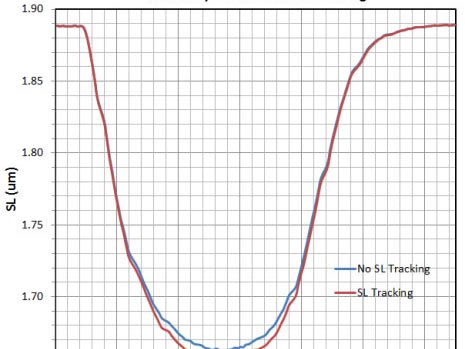
Can we quantify the effect of Dynamic Sarcomere Tracking?
Yes! The plot below shows sarcomere length calculated from the same contracting rat myocyte as is shown in the video. The ROI was identical for both measurements; the only difference was the use of Dynamic Sarcomere Tracking. Although the shape of the SL contraction is similar for both cases you will observe that the minimum sarcomere length is less when using tracking. We believe that this is due to averaging of more sarcomeres in the image when tracking is not used. Without tracking the number of sarcomeres in the calculation increases with decreasing sarcomere length. This has the effect of increasing the averaging which results in an underestimation of the true minimum. Another possible cause of the change in the minimum sarcomere length may be due to the translation of the tissue which results in a different region of sarcomeres being calculated when relaxed and when contracted.
![]()
Dynamic Tracking and Sarcomere Length Control
Our HVSL system is fast enough to be used for SL control during a contraction. The improved accuracy that is obtained by using the Dynamic Sarcomere Tracking should also improve SL control.
Please contact Aurora Scientific for further information about sarcomere measurement and Dynamic Sarcomere Tracking.


