1600A
Permeabilized Myocyte System – Microscope Mountable
The 1600A Permeabilized Myocyte Test System is designed to simplify the complex testing of contractile properties of skinned myocytes. A large cell mounting area provides ample room for cell attachment. Push button controlled motorized stages simplifies cell attachment to the high-speed servo motor (315D, 322D) and precision force transducer (400 series).
A temperature controlled 8-well bath plate allows for seamless transition of the myocyte between varying calcium concentrations. Direct mounting on an inverted microscope stage allows for observation and sarcomere spacing detection. An optional prism reticule can be included to provide accurate measurement of cell cross section.
With the 1600A, force, length and sarcomere spacing (with optional HVSL) can all be controlled and measured easily and repeatedly. This permits measurement of simple force-pCa to more complex mechanical characterization of the cell including kTr, length-tension, force-velocity and stiffness.
The system is truly turnkey, including components for data acquisition and analysis as well. Our Digital controller and Software Analysis package (600A) comes complete with a large library of standardized protocols to make the above characterizations straightforward. Choose the 1600A system for performance, precision and progress.

Inverted microscope with Permeabilized Myocyte Apparatus

Eight temperature controlled bath wells

Direct mounting of apparatus on inverted microscope
Stories of Success
Select References
- Payne et al. “The carbon monoxide prodrug oCOm-21 increases Ca2+ sensitivity of the cardiac myofilament” Physiological Reports (2024) DOI: 10.14814/phy2.15974
- Alegre-Cebollada, Jorge et al. “S-glutathionylation of cryptic cysteines enhances titin elasticity by blocking protein folding.” Cell (2014) DOI: 10.1016/j.cell.2014.01.056
- Krysiak et al. “Protein phosphatase 5 regulates titin phosphorylation and function at a sarcomere-associated mechanosensor complex in cardiomyocytes” Nature communications (2018) DOI: 10.1038/s41467-017-02483-3
- Herwig et al. “Protein Kinase D plays a crucial role in maintaining cardiac homeostasis by regulating post-translational modifications of myofilament proteins” International Journal of Molecular Sciences (2024) DOI: 10.3390/ijms25052790
- Hessel et al. “Myosin-binding protein C regulates the sarcomere lattice and stabilizes the OFF states of myosin heads” Nature Communications (2024) DOI: 10.1038/s41467-024-46957-7
- Gonçalves-Rodrigues et al. “In Vitro Assessment of Cardiac Function Using Skinned Cardiomyocytes” Journal of Visualized Experiments (2020) DOI: 10.3791/60427
- Pappritz et al. “Impact of syndecan-2-selected mesenchymal stromal cells on the early onset of diabetic cardiomyopathy in diabetic db/db mice” Frontiers in Cardiovascular Medicine (2021) DOI: 10.3389/fcvm.2021.632728
- Pappritz et al. “Impact of syndecan-2-selected mesenchymal stromal cells on the early onset of diabetic cardiomyopathy in diabetic db/db mice” Frontiers in Cardiovascular Medicine (2021) DOI: 10.3389/fcvm.2021.632728
- Kötter, Sebastian et al. “Human myocytes are protected from titin aggregation-induced stiffening by small heat shock proteins.” The Journal of Cell Biology (2014) DOI: 10.1083/jcb.201306077
- Horiguchi, Hiroshi et al. “Fabrication and evaluation of reconstructed cardiac tissue and its application to bio-actuated microdevices.” IEEE Transactions on NanoBioscience (2009) DOI: 10.1109/TNB.2009.2035282
System Components

315D/322D: High-Speed Length Controllers
The 315D/322D High-Speed Length Controllers give physiologists the ability to control and measure length of single cells, fibers and whole muscle with ease.
Learn More
400C: Force Transducers
The 400C series of force transducers is the next generation of our widely used 400A and 400B series. The 400C series enables contractile measurements from a variety of muscle types and sizes and are designed to meet the needs of muscle researchers.
Learn More
600A: Real-Time Muscle Data Acquisition and Analysis System
The 600A Digital Controller serves to integrate components and provide the researcher control of system operations, data collection and signal analysis.
Learn More
803B: Permeabilized Myocyte Apparatus – Microscope Mountable
Innovative 8–well plate designed for quick cell attachment and measurement of myocyte mechanical properties
Learn More
820A: Dual XYZ Motion Controller
Closed loop control of motorized stages to easily perform complex micro positioning with stunning precision
Learn More
901D: High-Speed Video Sarcomere Length
A simple, turn-key system for measuring sarcomere spacing in isolated muscle preparations.
Learn More
608C: LCD Monitor
A 22” widescreen LCD monitor providing high quality visualization of data collection
Explore Further

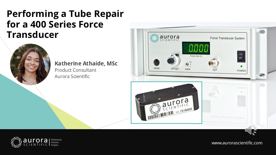
Calling in Reinforcements: Tube Repair of an ASI 400 Series Force Transducer
A step-by-step tech cast on how to perform a tube repair for Aurora Scientific's 400 Series Force Transducers, Katherine will share tips, tricks and best practices for best ...
Learn More
Writing Protocols with 600A for Permeabilized Tissues
This blog will provide a brief overview of how to write protocols using our Real-Time Muscle Data Acquisition and Analysis System (600A) software.
Learn More
Quick Start Guide to 600A
This blog will provide a brief overview of how to start-up and utilize our Real-Time Muscle Data Acquisition and Analysis System (600A) software.
Learn MoreResources
Parts & Accessories
| Model # | 1600A |
|---|---|
| 901D | The High-Speed Video Sarcomere Length (HVSL) system provides high-speed, accurate sarcomere length measurement from a live video image. Our 901D HVSL system uses a high-resolution, USB 3.0 camera to provide full-frame images (1280×1024) at up to 300 frames per second. This frame rate increases to 3,000 frames per second when the camera is windowed down to 16 pixel lines. Image capture and sarcomere length data can be controlled by our 600A Digital Controller software which runs on a real-time Linux operating system. |
| 830A | Half-rack adapter that allows half-rack instruments to be mounted in standard 19” racking. Works with models: 315D/322D, 400C series, 701C and 825B. |
| 831A | Heavy gauge, steel desktop or shelf-top rack, with standard 19” width for any Aurora Scientific Instruments. Available in various heights. (6U high for 1200A and 1300A, 5U high for 1400A, 1500A and 1600A systems). |
