Courtesy of Glass et al. (2019).
The larynx is an organ that sits in the top of the neck and holds the vocal cords. It contains several muscles that work together to move the vocal cords and cartilage and produce vocalizations. Mammals, reptiles, and amphibians all have a larynx, while the vocal organ within birds is the syrinx. Recent studies are helping elucidate how muscles within this organ change over the course of development and during disease pathogenesis.
Increasing Muscle Speed Drives Changes in the Neuromuscular Transform of Motor Commands During Postnatal Development in Songbirds
Adam et al. (2020) analyze the effect of development on motor control within the songbird syrinx. It is clear that neural pathways change over the course of song learning, but less is known about how developing muscles influence changes in sound. The authors hypothesized that syringeal muscle biomechanical properties change over the course of vocal development in songbirds. They isolated muscle fiber bundles from the syrinx of zebra finches, attached them to our 400A force transducer, and stimulated them with our 701C high-powered stimulator in order to measure muscle contraction speed. They found that over the course of development, syringeal muscle speed increased in male songbirds but not female ones. At faster speeds, muscles produce increasingly higher force, and supralinear summation occurs. The authors also showed that faster muscles produced higher spike timing sensitivity. This study shows that developmental changes in songbird vocal muscles lead to differences in how neural signals are translated into force profiles, which changes the sound of the bird’s songs.
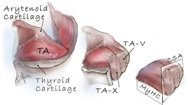
Laryngeal Muscle Biology in the Pink1-/- Rat Model of Parkinson Disease
Glass et al. (2019) analyzed changes in laryngeal muscles in early Parkinson disease. This condition leads to pathological changes in the nerves and muscles of the larynx and pharynx, causing defects in vocal communication. The authors hypothesized that these changes emerge in early disease stages, before motor symptoms and other disease signs. To explore this hypothesis, the authors used a rat model of Parkinson disease in which the Pink1 gene is knocked out, leading to deficits in motor function, vocalizations, and swallowing. The researchers isolated the thyroarytenoid (TA) muscle, which forms the bulk of the vocal cord, and attached it to our 305C dual-mode muscle lever to measure the muscle’s contractile properties. Compared with wild-type control rats, Pink1-/- rats had reduced force levels during premanifest disease stages. The Parkinson disease rat model also exhibited increased levels of myosin heavy chain isoform (MyHC) in the TA, higher numbers of centrally nucleated myofibers, and smaller myofibers in the vocalis region of the TA. This report shows that vocal impairments occur early on in the course of Parkinson disease and may be used as a way to detect the condition early.


