Coming off the heels of this year’s Basic Cardiovascular Sciences Scientific Sessions (BCVS 2024), the following publication review focuses on an array of cardiac research, ranging from human induced pluripotent stem cells (hiPSC) to whole animal studies. Paralleling the meeting’s breadth of cardiac focuses, this review will cover recent insights on the development of hiPSC–derived cardiomyocytes (hiPSC-CMs), the sex-dependent progression of heart failure, and the regulation of t-tubule structure and function.
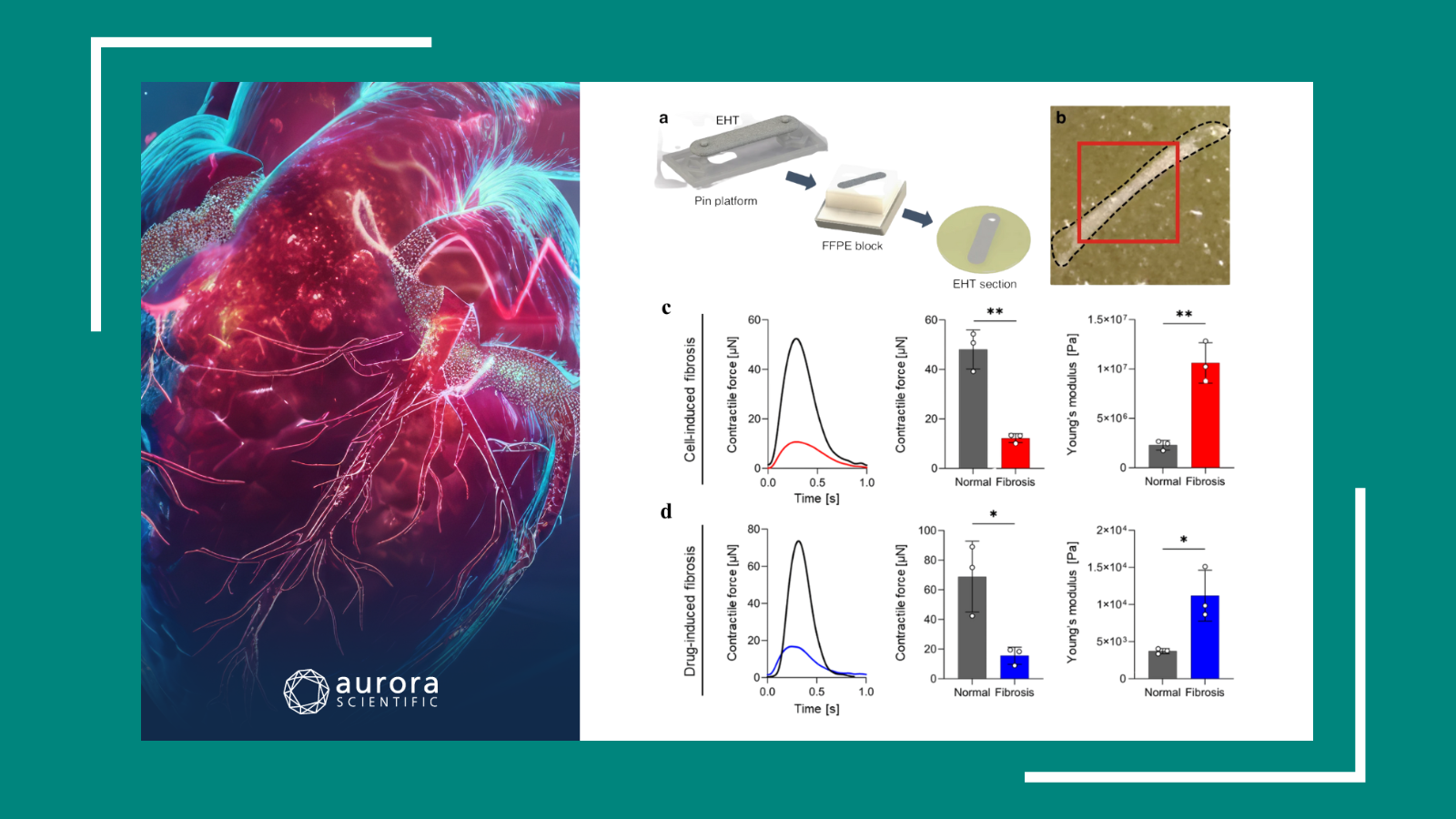
Featured image (adapted from © Rossler et al. (2024), licensed under CC BY 4.0 DEED) demonstrating A) three-dimensional representations of hiPSC-ECTs from lactate and MACS purification methods. B) α-Actinin and DAPI immunofluorescence labeling on lactate and MACS hiPSC-ECTs. C) Sarcomere length comparison. D) Raw twitch force (TF) and E) normalized averaged TF (NForce) curves for lactate (red) and MACS (blue) hiPSC-ECTs.
Lactate- and immunomagnetic-purified hiPSC–derived cardiomyocytes generate comparable engineered cardiac tissue constructs
Human induced pluripotent stem cells (hiPSCs) are a powerful research tool, enabling disease modelling, drug discovery, and precision therapies. The ability to differentiate hiPSCs into virtually any cell type of interest has infiltrated the fields of neuroscience, nephrology, gastroenterology, and cardiology, showcasing their far-reaching potential across scientific disciplines. Within the cardiac field, hiPSC-derived cardiomyocytes (hiPSC-CMs) have been strategically employed to model cardiac diseases and predict drug-induced cardiotoxicity. Despite these advances, concerns have been raised surrounding the purification methods of hiPSC-CMs during the generation of 3D engineered cardiac tissue (hiPSC-ECT). While lactate purification is a popular and cost-effective methodology, a recent study by Davis et al. (2022) implicated the technique in an ischemic cardiomyopathy-like phenotype compared to magnetic antibody-based cell sorting (MACS) purification. To address this, Rossler et al. (2024) performed a detailed analysis of the structure, function, and proteome between lactate and MACS hiPSC-ECTs.
Each hiPSC-ECT construct was attached to the 801B Small Intact Muscle Apparatus using sutures, and the 701C High-Power Stimulator paced the tissues at 1 Hz. Isometric twitch forces were recorded using the force readouts from the 403A Force Transducer, as the constructs were incrementally stretched to optimal length. After further functional analyses, including the assessment of Ca2+ transients and sarcomere length, the constructs were flash frozen for proteomic analysis.
Across both types of constructs, the assessment of isometric twitch force and Ca2+ transient measurements revealed similar functional performances. Moreover, structural and proteomics-based analyses revealed that there were no significant differences in sarcomere length, protein pathway expression, or myofilament proteoforms. As such, these comprehensive assessments suggest that lactate purification does not result in a notable difference from MACS purification when hiPSC-CMs are subsequently grown in ECTs.
Sex‑specific cardiovascular remodeling leads to a divergent sex‑dependent development of heart failure in aged hypertensive rats
The rising prevalence of heart failure with preserved ejection fraction (HFpEF) underscores a critical need for increased research efforts within the field. In fact, several emerging studies point to sex differences in heart failure (HF), including variations in risk factors, pathophysiology, and response to therapy. Despite these findings, females are still under-represented in both experimental and clinical studies, leading to significant gaps in our understanding of the disease. To bridge this gap, Kovács et al. (2024) employed 15-week- and 1-year-old female and male hypertensive transgenic rats for a crucial comparison of cardiac function.
Using the 1600A Permeabilized Myocyte Test System, single cardiomyocytes from each of the transgenic and control groups were selected under an inverted microscope, attached with silicon adhesive to the 315C High-Speed Length Controller, and assessed with the 403A Force Transducer. Ca2+ sensitivity of the skinned cardiomyocytes was measured by transferring the preparations from relaxing to activating solutions across the wells of the apparatus.
This investigation into cellular cardiac function revealed sex-dependent differences in Ca2+-dependent force production in both wildtype and transgenic rats. Specifically, males had increased pCa50 values, while females had decreased values compared to WT controls. Moreover, transgenic females exhibited decreased Fmax values compared to controls. Beyond the cellular level, transgenic male rats showed significantly higher mortality at 1 year than any other group, as well as cardiac enlargement and LV hypertrophy. At the protein level, phosphorylation of cMyBP-C and cTnI, were higher in WT males compared to WT females, and further increased in TG males. Taken together, these findings provide further evidence of sex-specific cardiovascular remodeling in the development of HF. The outlined sex differences in cardiac dysfunction further our understanding of HF development across males and females and importantly pave the way for more tailored therapies and treatments.
Regulation of cardiomyocyte t-tubule structure by preload and afterload: Roles in cardiac compensation and decompensation
T-tubules are essentially membrane invaginations that play a critical role in triggering cardiomyocyte contraction by facilitating the transmission of action potentials. In fact, disruptions in t-tubule structure or regulation can impair cardiac performance, resulting in diseases such as heart failure. Although it is known that high workload during heart failure is associated with a disruption in cardiomyocyte t-tubules as well as Ca2+ homeostasis, whether changes in preload and afterload may promote adaptive t-tubule remodeling remains unknown. Therefore, Ruud et al. (2024) sought to uncover how changes in mechanical load affect t-tubule integrity and cardiac function.
Papillary muscles were harvested from the hearts of 9–12-week-old male Wistar rats, and mounted on the 1205A Isolated Muscle System, with one end of the muscle attached to the 300B-LR Dual-Mode Lever System. The degree of afterload was varied by the rigidity of the lever arm, and custom-made software in LabView was used to control the lever arm and acquire data.
Upon analysis of the isolated papillary muscles there was a clear bell-shaped relationship between cardiomyocyte t-tubule density and changes in preload or afterload. Further analyses with myocardial samples from rodent and human hearts revealed that moderate increases in load led to t-tubule proliferation, enhanced calcium handling, and improved cardiac function. However, upon excessive load, there is a significant reduction in t-tubule density, and a progression to heart failure with reduced ejection fraction (HFrEF). As such, this study demonstrates that systolic tension is vital in regulating t-tubule density and cardiac function under both normal and pathological conditions.
Conclusions
Recent advances in the cardiac field are driving our understanding of heart disease modelling, pathogenesis, and novel therapeutic avenues. Through our improved knowledge of 3D engineered cardiac tissue generation, sex differences in heart failure, and the effect of systolic tension on cardiac function, these studies by Rossler et al. (2024), Kovács et al. (2024), and Ruud et al. (2024) collectively echo a promising trend of novel insights.