The analysis of cardiac tissue function is vital in the research of cardiovascular diseases and disorders, with researchers relying on innovative tools and technologies that offer unparalleled precision and reliability. Aurora Scientific’s precision research tools have been crucial in enabling key advancements in this field. Here, we delve into three studies which have harnessed the power of Aurora Scientific’s research tools to enable unique approaches to cardiac physiology and cardiomyocyte function.
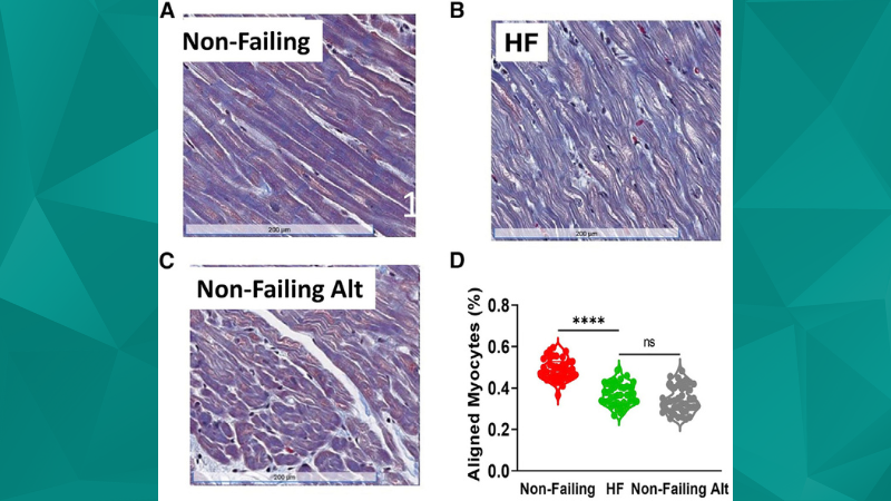
Featured image (©Ma et al. (2022), licensed under CC BY), shows human cardiomyocyte alignment by histology analysis from Non-Failing and patients with HF (HFrEF).
Myofibril Orientation as a Metric for Characterizing Heart Disease
Heart disease is typically diagnosed and monitored through assessing large-scale characteristics, such as heart size, weight, and blood flow. Yet, a critical unknown area is the microscopic changes in the heart’s architecture, specifically the orientation of myofibrils – the basic contractile units of muscle cells – and its relationship with heart disease. This gap in knowledge spurred researchers in this article by Ma et al. (2022) to explore whether myofibril orientation could serve as a novel metric for characterizing heart disease.
To understand the orientation of these miniature muscular structures, researchers employed Aurora Scientific’s 402A Force Transducer and Muscle Dynamic Control System. These tools were used to analyze isolated cardiomyocyte bundles under various conditions, simulating both healthy and diseased states. Aurora Scientific’s tools allowed for meticulous control and accurate force measurements, a prerequisite for such intricate studies. X-ray diffraction patterns collected at various sarcomere lengths revealed distinct changes in myofibril orientation in disease-imitating conditions.
This research provides a microscopic view into heart disease and opens up a novel frontier for diagnostic techniques. The ability to detect disease at a micro-architectural level could enhance diagnostic accuracy, allowing clinicians to detect heart diseases earlier and tailor treatments more effectively.
Subcellular Remodeling in Filamin C Deficient Mouse Hearts Impairs Myocyte Tension Development during Progression of Dilated Cardiomyopathy
While the role of genetics in heart diseases is established, the specific mechanisms and pathways are not fully understood. For instance, the exact role and implications of Filamin C deficiency, a protein vital for cellular structure, remains largely unexplored. This study by Powers et al. (2022) aimed to elucidate the impact of Filamin C deficiency on heart tissue structure and function, potentially revealing new insights into genetic influences on heart disease.
Following dissection of small intact papillary muscles from the right ventricle, Aurora Scientific’s 1500A Small Intact Muscle System allowed researchers to measure force dynamics under Filamin C-deficient conditions. They were able to detect and analyze the structural and functional changes in cardiac tissues, which revealed a significant impairment in heart muscle tension development due to Filamin C deficiency.
This research is pivotal as it unravels the impacts of Filamin C deficiency on heart function at the cellular level, bridging the gap between genetic influences and heart diseases. These insights could potentially lead to new gene-targeted therapies for cardiac conditions. Furthermore, the findings underscore the importance of precise, high-quality research tools, like those from Aurora Scientific, in understanding complex biological processes.
Epicardially Secreted Fibronectin Drives Cardiomyocyte Maturation in 3D-Engineered Heart Tissues
Regenerative medicine has seen considerable advancements in recent years, with 3D-engineered heart tissues (3D-EHTs) playing an increasingly crucial role. However, the factors that drive the maturation of cardiomyocytes, the fundamental cells in these tissues, remain unclear. This study by Ong et al. (2023) aimed to investigate the role of fibronectin, a protein secreted by the outermost layer of the heart (epicardium), in driving cardiomyocyte maturation in 3D-EHTs.
Researchers used Aurora Scientific’s 400A force transducer and 312B Length Controller to conduct precise Frank-Starling force measurements on the 3D-EHTs, enabling them to examine both passive tension and active force dynamics. Detailed analyses revealed that the epicardially secreted fibronectin significantly influenced the maturation and functionality of cardiomyocytes within the 3D-EHTs, an observation not previously documented.
This study marks a significant stride in cardiac regenerative medicine, suggesting that modulating fibronectin secretion could enhance the development of 3D-EHTs. Such advancements could have significant implications for treating heart failure and other cardiovascular diseases in the future.
Conclusion
These three studies exemplify how Aurora Scientific’s precision research tools can catalyze advancements in understanding heart diseases. The studies not only underscore the necessity of such equipment for high-quality cardiac research but also illustrate their potential to guide the future of cardiovascular diagnostics and therapy development. The valuable insights gained in these studies open new horizons for improved patient outcomes, highlighting the significance of technological advancements in the biomedical research landscape.


