Physiology is a far-reaching discipline, encompassing everything from cell function to whole-body function. Beyond underpinning translational and clinical medicine, physiology research informs all aspects of our health and well-being. In recognition of this expansive field, physiologists across the world will gather for the American Physiology Summit, a popular venue to network with colleagues, learn from attendees, and share their research. In the spirit of the conference, the following publication review covers forthcoming insights in muscle physiology, including smooth muscle mechanics, a neutralizing treatment for mitochondrial myopathies, and the role of Myc in skeletal muscle growth.
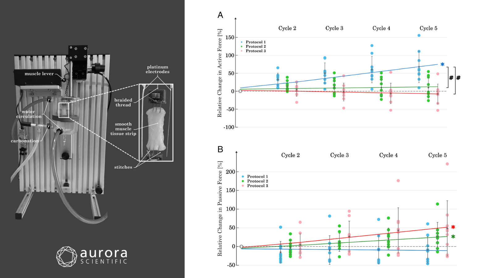
Featured image (adapted from ©Kiem et al. (2025), licensed under CC BY 4.0) depicting the smooth muscle mechanics setup on the 800A: In-vitro Muscle Apparatus (left). On the right are the resultant change in active (A) and passive (B) forces at λ = 2 over 5 cycles for each of the three protocols.
Reproducibility of smooth muscle mechanical properties in consecutive stretch and activation protocols
Organ models serve as important preclinical test systems and are critically dependent on input data. This data is obtained through repeated mechanical experiments to provide an accurate model of the representative organ. In the instance of smooth muscle models, acquiring this input data is particularly challenging, due to rapid changes in active and passive properties during measurement. To address this challenge, Kiem et al. (2025) sought to reproduce the initial mechanical properties of SM tissue after consecutive stretch and activation protocols.
Urinary bladder tissue strips from nine healthy, female domestic pigs were sewn into 3D-printed mounting parts and attached vertically to Aurora Scientific’s 305C-LR Dual-Mode Lever System, which was secured on the 800A: In-vitro Muscle Apparatus. The length and force signals were recorded using the 604A: Data Acquisition Signal Interface, and data was acquired on the 610A: Dynamic Muscle Control software. Each protocol included five repetitive experimental cycles with stimulations at a long muscle length and a varied number of stimulations at a short muscle length (Protocol 1: 0 stimulations, Protocol 2: 1 stimulation, Protocol 3: 2 stimulations).
Upon analysis, Protocols 2 and 3 successfully reproduced the initial active force at long length over five cycles but did not maintain the initial passive forces. Conversely, Protocol 1 induced increasing active forces with each cycle, and was most effective in maintaining constant passive force over the cycles. Taken together, this study presents a new method for obtaining reproducible smooth muscle parameters and provides a foundation for future studies exploring smooth muscle mechanical properties.
GDF15 Neutralization Ameliorates Muscle Atrophy and Exercise Intolerance in a Mouse Model of Mitochondrial Myopathy
Primary mitochondrial myopathies (PMMs) are genetic disorders caused by mutations that perturb mitochondrial function. PMMs can lead to a constellation of symptoms but predominantly impact skeletal muscle through the characterized loss of muscle mass and strength, exercise intolerance, and fatigue. Intriguingly, both patients with PMM and mouse models of mitochondrial myopathy exhibit elevated growth differentiation factor 15 (GDF15) levels, suggesting it may play a key role in the disease process. To explore the therapeutic potential of GDF15 neutralization, Flaherty III et al. (2025) assessed whether treatment with a GDF15 neutralizing antibody could alleviate muscle impairments in mitochondrial DNA polymerase gamma (POLG) mutator mice, a mouse model of PMM.
POLG mutator, anti-GDF15 antibody treated (POLG-GDF15 mAB2), and wildtype mice underwent assessments for voluntary wheel running, treadmill running, and in-vivo muscle function. Aurora Scientific’s 300C-FP Dual-Mode Lever System and 701C High-Power Stimulator were used in conjunction to assess in-vivo plantar flexion, and the 615A: DMC and DMA Software package was used for data collection and analysis.
When measured, circulating GDF15 levels were elevated in POLG mice as early as 3 months of age, with a marked surge by 6 and 10 months. Alongside this biochemical shift, the mice began to present clear signs of physical decline: stunted weight gain from around 6 months, noticeable reductions in both fat and lean body mass, diminished plantar flexors force, and a drop in voluntary wheel running activity. Strikingly, neutralizing GDF15 reversed many of these phenotypes, leading to significant weight gain, preserved fat mass, and importantly, restored muscle function and exercise tolerance. These improvements highlight GDF15 neutralization as a powerful therapeutic strategy from which to improve physical performance and mitigate symptoms in PMM patients.
Muscle fiber Myc is dispensable for muscle growth and its forced expression severely perturbs homeostasis
Myc is an oncogenic transcription factor that regulates the genetic expression of cell growth, proliferation and differentiation. Although Myc expression is low in adult skeletal muscles, it is known to increase after hypertrophic resistance exercise, implicating a potential involvement in skeletal muscle growth (hypertrophy). To test this, Ham et al. (2025) interrogated both Myc overexpression and elimination in skeletal muscle fibers and muscle stem cells (MuSC).
In a comprehensive assessment, a combination of functional and molecular experiments were performed, including in-vitro muscle force measurements, histology, immunostaining, digital PCR, and RT-qPCR. For the former, fast-twitch extensor digitorum longus (EDL) and slow-twitch soleus muscles were carefully isolated, sutured, and mounted on Aurora Scientific’s 1200A Isolated Muscle Test System for Rodents. After defining optimal length, muscles underwent tetanic, force-frequency, and fatigue protocols.
Despite reduced Myc expression, mice with eliminated Myc in their skeletal muscle fibers showed no major differences in muscle mass or isolated muscle force compared to controls. In fact, they exhibited only a slight reduction in peak twitch force in male EDL muscles and had overall preserved muscle function and fatigability. These findings suggest that muscle fiber Myc knockout has minimal impact on normal muscle development and performance. Although Myc was found to be essential in muscle stem cells for regeneration, muscle fiber Myc was surprisingly unnecessary for postnatal or induced growth—and when overexpressed, disrupted muscle structure and function. These findings overturn the idea of Myc as a straightforward growth promoter in muscle, challenging its assumed role as a driver of muscle hypertrophy.
Conclusions
These studies by Kiem et al. (2025), Flaherty III et al. (2025), and Ham et al. (2025) highlight the diverse and evolving landscape of muscle physiology research – from uncovering smooth muscle mechanics and therapeutic interventions to challenging assumptions about growth regulators. Taken together, they underscore the importance of integrative research in shaping our understanding of muscle health, function, and therapeutic potential.


