Tip 3: Glue can be used to Attach a Skinned Myocyte to an ASI Force Transducer and Length Controller
Depending on the size of the tissue preparation it is possible to attach muscle tissue to a length controller and force transducer using glue. Typically the glue method is used with very small preps including skinned myocytes.
In this method a small amount of glue is placed on the tip of micropipettes attached to a force transducer and to a length controller. The tissue is then brought into contact with the glue and the myocyte is attached.
Two main types of glue are commonly used. The first is an expanding foam called GREAT STUFF manufactured by The Dow Chemical Company. It is readily available in the USA in stores such as Home Depot. The other is a silicone based glue manufactured by Dow Corning Corp.
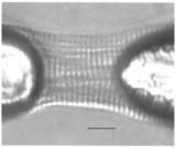
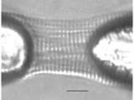
Figure 1 Myocyte Glued to Micropipettes
The glue method of attaching a skinned myocyte to a force transducer and high speed length controller is explained well in the following paper by Sweitzer and Moss.
The method consists of forming micropipettes with a tip diameter of 4 microns and attaching these micropipettes to the force transducer and lever arm using paraffin wax, shellac or dental impression compound. A drop of glue is placed on a microscope slide or cover slip along with a drop of cell suspension. An appropriate myocyte is chosen by viewing the suspension droplet with a microscope. The two micropipette tips are then dipped into the glue and lowered onto each end of the myocyte. The glue is left to cure for about 45 minutes and then the pipettes are translated along with the suspended myocyte to the test chamber. Aurora Scientific’s 803B Skinned Myocyte Test Apparatus was designed for exactly this experiment and it provides a large cell mounting and gluing area along with 8 test wells for different calcium concentrations. Force-pCa data can be obtained with this system.
P. de Tombe, Loyola University, Chicago and G. Stienen, VU University Medical Centre, Amsterdam are two researchers using glue to attach skinned myocytes to an Aurora Scientific high speed length controller and force transducer.


