As December draws to a close, it brings the familiar mix of reflection and anticipation, with the year winding down and researchers pausing to take stock of the scientific harvest. Over the twists and turns of 2025, the research landscape has been alive with innovation across neuroscience, tissue engineering, and cardiovascular technology, offering fresh insights into how the body senses, builds, and sustains itself. From circuits that prime our appetite before the first bite, to acoustically assembled tissues that flex and fire like native muscle, and implantable cardiac sensors that keep the heart under vigilant watch, this year has delivered discoveries as clever as they are impactful. Continuing our annual end-of-year tradition, this year’s final publication review spotlights the standout studies that captured the imagination of the field, pushing the boundaries of sensory biology, regenerative medicine, and patient-specific cardiac monitoring.
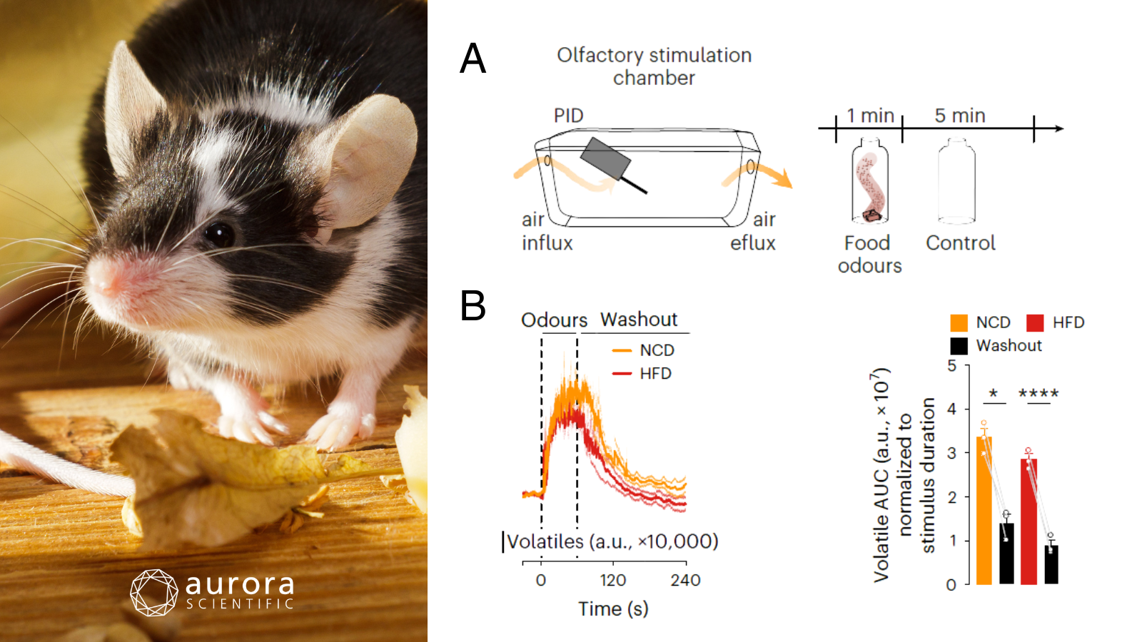
Featured image (photo by Shaiith from Getty Images, via Canva.com with figures adapted from ©Bulk et al. (2025), licensed under CC BY 4.0) shows a sniffing mouse and the experimental setup. A) Schematic of the olfactory stimulation chamber, which delivers controlled odourized air via an olfactometer and Aurora Scientific’s 200C: miniPID Fast Response Olfaction Sensor for volatile concentration measurements, B) Volatile concentrations during 1 min of food odour exposure (NCD, orange; HFD, red) followed by 5 min of control odour (empty bottle; black; time curve: n = 3 trials; AUC: repeated-measures one-way ANOVA; n = 3 trials; P = 0.0002, P = 0.001, a.u. denotes arbitrary units).
A food-sensitive olfactory circuit drives anticipatory satiety
Food sensory cues, particularly olfactory signals, are known to trigger anticipatory (cephalic-phase) responses that prepare the body for nutrient intake and can rapidly modulate the activity of key feeding-related neurons. However, despite evidence that food cues influence metabolic and hypothalamic circuits, the specific behavioural relevance of olfactory inputs for feeding regulation and the underlying neural pathways remain poorly defined, especially in the context of obesity. Bulk et al. (2025) sought to address this gap by identifying and functionally characterizing a previously unrecognized olfactory bulb–to–medial septum circuit that integrates food odours to prime anticipatory satiety before eating. By combining whole-brain mapping, in-vivo neuronal recordings, and circuit-specific manipulations in lean and obese mice, they demonstrate how olfactory-driven neural activity shapes food intake and how this mechanism is disrupted in diet-induced obesity.
To examine how food odours engage olfactory–septal circuits, a combination of genetic mouse models, viral tracing, chemogenetics, optogenetics, and in-vivo calcium imaging was used alongside behavioural and metabolic assays under control and high-fat diet conditions. Neuronal activity was recorded during food perception, controlled odour exposure, and hormonal manipulations using fibre photometry, while circuit function was tested through targeted stimulation paired with feeding and behavioural measurements. To precisely control and quantify olfactory stimuli, a custom-built olfactory stimulation chamber was coupled with Aurora Scientific’s 200C: miniPID Fast Response Olfaction Sensor, which provided real-time measurement of volatile concentrations during odour delivery.
Across unbiased whole-brain mapping approaches, food odour exposure activated classical olfactory regions and feeding-related hypothalamic nuclei, while uniquely and selectively engaging the medial septum (MS), where glutamatergic MSVGLUT2 neurons showed robust and specific responses to food cues. These neurons displayed a biphasic activity pattern (rapid activation during food odour perception followed by sustained inhibition after ingestion) and received direct monosynaptic input from olfactory bulb mitral and tufted cells, forming a circuit that constrained food intake when acutely stimulated before feeding in lean mice. Rapid, reproducible increases in volatile concentrations tightly matched neuronal activation and sniffing behaviour, confirming that MSVGLUT2 responses were driven by food odours rather than generalized olfactory or arousal cues. Collectively, these findings identify a previously unrecognized olfactory–septal circuit that encodes food odour perception to promote anticipatory satiety, a mechanism that is disrupted in diet-induced obesity and highlights how sensory processing can directly shape feeding behaviour.
Acoustofluidic bioassembly induced morphogenesis for therapeutic tissue fabrication
The fabrication of functional in-vitro tissues has long aimed to recapitulate the orderly spatial organization of cells that emerges during morphogenesis in-vivo, as such architecture is essential for tissue maturation and function. Despite advances in bioprinting and scaffold-based approaches, it remains challenging to simultaneously control cellular alignment, dense aggregation, and multi-scale organization without compromising cell viability or resolution. Therefore, Kang et al.(2025) introduced an acoustofluidic bioassembly–induced morphogenesis strategy that harnesses acoustic forces to precisely arrange cells prior to differentiation. By coupling this controlled bioassembly with subsequent in-vitro and in-vivo maturation, the study demonstrates a scalable platform capable of generating structurally and functionally advanced therapeutic tissues.
Piezoelectric acoustofluidic devices (systems that use electrically generated sound waves) were engineered to organize cells within hydrogels as they solidified, enabling precise control over tissue architecture. Mouse fibroblasts were reprogrammed into induced myogenic progenitor cells (iMPCs) and combined with motor neurons, then assembled into aligned neuromuscular tissues using fibrin- or GelMA-based hydrogels. Tissue maturation was evaluated using imaging, calcium signaling, gene expression, and electrophysiological measurements before transplantation into a mouse model of volumetric muscle loss. Muscle function was quantitatively assessed in-vivo using Aurora Scientific’s 1300A: Whole Animal Muscle Test System and 615A: Dynamic Muscle Control and Analysis Software.
Acoustofluidic bioassembly (AB) enabled the rapid fabrication of neuromuscular tissues with highly organized cellular architecture, promoting direct interactions between iMPCs and primary motor neurons (pMNs). Tissues generated with AB exhibited enhanced maturation, evidenced by increased myofiber diameter, fusion index, striation, NMJ formation, and synchronized calcium transients compared to randomly distributed controls. Functional assessments demonstrated that transplanted iMPC+MN AB tissues restored muscle contractile force, contraction and relaxation rates, and fatigue resistance, closely approaching normal muscle performance. These results highlight AB as a versatile approach for engineering structurally and functionally advanced neuromuscular tissues with strong regenerative potential in volumetric muscle loss.
VITALS: an implantable sensor network for postoperative cardiac monitoring in heart failure patients
Heart failure poses a formidable challenge to cardiovascular care, with high-risk patients experiencing significant postoperative morbidity and mortality despite advances in surgical interventions. Traditional follow-up assessments are intermittent and often fail to detect early cardiac deterioration, leaving a critical gap in post-acute monitoring. While myocardial strain has emerged as a sensitive early indicator of cardiac dysfunction, current clinical methods are limited in their ability to provide continuous, remote measurements. To address these limitations, Kight et al. (2025) investigated whether an implantable, epicardial sensing network could enable real-time, multi-axial strain monitoring, offering the potential for proactive, patient-specific interventions.
To evaluate VITALS (Ventricular Implantable Technology for Accurate Live Strain) sensor performance, micro-electromechanical systems (MEMS)-based barometer sensors were fabricated on flexible printed circuit boards (PCBs) and encapsulated in silicone for durability and flexibility. These sensors were calibrated on a 3D-printed jig by dynamically stretching each element up to 5 mm using Aurora Scientific’s 300C: Dual-Mode Lever. Benchtop studies with heart-shaped silicone phantoms and in-vivo studies in anesthetized pigs assessed sensor performance under physiological and pathological conditions, while echocardiography and pressure-volume (PV) loop catheters were used in parallel to validate strain and volume measurements, with careful synchronization of sensor and imaging data.
The VITALS sensors were found to accurately capture cardiac mechanics across benchtop and in-vivo conditions, despite a few notable challenges. Mainly, the low-stiffness gel encapsulation reduced sensitivity to shear forces, allowing the sensors to maintain signal fidelity even under simulated tissue adhesion, while calibration confirmed precise mapping of barometer readings to mechanical strain. In-vivo, the sensor network tracked biventricular strain, left ventricular volumes, and aortic pressure with strong agreement to clinical gold-standard metrics, even during ischemia and pharmacologically induced changes in contractility. Taken together, these findings demonstrate the robustness and clinical relevance of the VITALS system, providing a foundation for real-time cardiac monitoring in complex physiological and pathological states.
Conclusions
These studies by Bulk et al. (2025), Kang et al.(2025), and Kight et al. (2025) illuminate how the body senses, builds, and sustains itself, from olfactory circuits that prime appetite, to acoustically patterned tissues that replicate native muscle function, and implantable sensors that provide real-time cardiac vigilance. Collectively, they highlight the power of integrating sensory, structural, and monitoring systems, offering a glimpse into the innovative approaches shaping the future of physiology, regenerative medicine, and patient-specific care as we step into the New Year.
Follow Aurora Scientific’s journey over the years: Checkout out last year’s end-of-year publication review, ‘Best of 2024: Intricate Advancements’.


