Cancer cachexia is a complex, treatment-resistant syndrome marked by muscle wasting, weight loss, and metabolic disruption, impacting the majority of patients with advanced cancer. As a major driver of poor outcomes, it remains an urgent and under-addressed challenge. In the lead-up to the 8th Cancer Cachexia Conference in Turin, Italy, this review spotlights three compelling studies that push the field forward: the sex-specific role of myofiber-derived FoxP1 in muscle loss, the ability of mitochondrial-targeted SkQ1 to preserve strength independent of atrophy in ovarian cancer, and a novel head and neck cancer model capturing the systemic toll of cachexia. Muscling our way through 2025, these recent advances flex new insights in cancer cachexia.
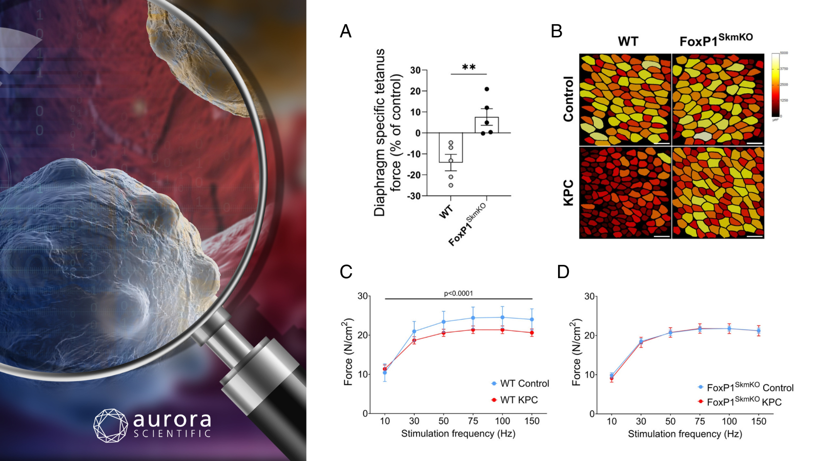
Featured image created with 3D software of a cancer cell (©Vitanovsky/Getty Images via Canva.com) and adapted figures from ©Schonk et al. (2025) (licensed under CC BY 4.0), displaying ex-vivo diaphragm strip assessments in wildtype (WT) and FoxP1SkmKO mice injected with either KPC cancer cells or saline. A) Specific tetanic force measurements; B) Representative diaphragm muscle sections with fibers color-coded by cross-sectional area (CSA), scale bar = 100 µm; C) Force-frequency curves for diaphragm strips from WT mice; and D) FoxP1SkmKO mice.
Role of myofiber-specific FoxP1 in pancreatic cancer-induced muscle wasting
Cancer cachexia is a life-threatening metabolic syndrome marked by progressive muscle wasting and weight loss, contributing to nearly a third of all cancer-related deaths. Despite its severe impact on survival and quality of life, no approved therapies exist, largely due to the complexity of its underlying mechanisms. Prior work identified the transcriptional repressor FoxP1 as elevated in cachectic muscle, but its functional role remains unclear. To address this, Schonk et al. (2025) generated myofiber-specific FoxP1 knockout mice to investigate how this gene influences muscle physiology and cancer-induced wasting, with attention to potential sex-specific effects.
To investigate the role of FoxP1 in cancer-induced muscle wasting, researchers used myofiber-specific FoxP1 knockout (FoxP1SkmKO) mice and an orthotopic pancreatic cancer model to induce cachexia. Muscle function was evaluated ex-vivo using Aurora Scientific’s 300C Dual-Mode Muscle Lever, which allowed precise measurement of diaphragm contractile strength under controlled conditions. Additional analyses included histology for muscle fiber size and composition, and molecular techniques like RT-qPCR to assess gene expression. Together, these methods enabled a comprehensive assessment of muscle structure, function, and molecular changes in response to FoxP1 deletion during cachexia.
Myofiber-specific deletion of FoxP1 protected male mice from pancreatic cancer-induced muscle wasting and weakness. Specifically, WT tumor-bearing mice showed a 14% drop in diaphragm-specific tetanic force and reduced force-frequency responsiveness, both of which were prevented in FoxP1SkmKO mice. These mice also retained muscle mass and fiber size compared to WT counterparts, despite similar tumor burdens. In contrast, female FoxP1SkmKO mice were not protected against cancer-induced muscle atrophy, underscoring a striking sex-specific difference in FoxP1’s role during cachexia. These findings highlight FoxP1 as a key driver of cancer-induced muscle dysfunction in males and a potential therapeutic target for combatting cachexia-associated muscle weakness.
Mitochondrial-targeted plastoquinone therapy prevents early onset muscle weakness that occurs before atrophy during ovarian cancer
Cancer cachexia is a debilitating syndrome marked by progressive muscle wasting and weakness, severely impacting quality of life, treatment tolerance, and survival. Despite decades of research, no effective therapies exist, in no small part due to limitations in current animal models that poorly mimic human disease. While most studies focus on muscle atrophy, emerging evidence suggests that muscle weakness actually precedes atrophy and may arise from early mitochondrial dysfunction. Therefore, Delfinis et al. (2025) aimed to test whether targeting mitochondrial stress with the antioxidant SkQ1 could prevent both early and late muscle weakness in a novel, translational model of metastatic ovarian cancer cachexia.
Female mice were injected with ovarian cancer cells to develop a clinically relevant, orthotopic model of metastatic ovarian cancer cachexia. The mitochondrial-targeted antioxidant SkQ1 was administered via drinking water to evaluate its impact on muscle weakness and atrophy at both early and late stages of disease. Muscle function was assessed using Aurora Scientific’s 1300B: Integrated Whole Animal System, configured with the 305C Dual-Mode Muscle Lever which allowed precise measurements of in-situ and ex-vivo force production in different muscle types (tibialis anterior, diaphragm, and FDB). This enabled high-resolution force-frequency and fatigue testing, revealing how SkQ1 modulates muscle strength independently of muscle size.
SkQ1 treatment partially preserved muscle function in a mouse model of ovarian cancer-induced cachexia. Notably, SkQ1 improved force production in both the tibialis anterior (TA) and diaphragm during early-stage disease, preventing 46% and 62% of cancer-induced muscle weakness, respectively, without altering muscle size. At late-stage disease, SkQ1 still enhanced diaphragm force output by 30–41% and fully preserved force during high-frequency stimulation in the FDB, in part by improving calcium handling. These findings highlight SkQ1’s potential to selectively counteract cancer-induced muscle weakness through mechanisms independent of muscle atrophy, offering a promising therapeutic avenue for preserving functional capacity in cancer cachexia.
B0092 tumor-bearing mice are a new model for the study of cachexia in head and neck cancer
Head and neck cancer (HNC) affects about 4% of all cancer patients but contributes to significant mortality and morbidity, especially due to cachexia. Despite its prevalence, the mechanisms behind HNC-induced cachexia remain poorly understood, largely due to the lack of suitable animal models. Livingston et al. (2025) aimed to address this by developing and characterizing a novel preclinical model using tobacco-induced B0092 oral squamous cell carcinoma in immunocompetent mice.
Muscle function was assessed ex-vivo using Aurora Scientific’s 300C Dual-Mode Muscle Lever, which measured contractile force of isolated extensor digitorum longus (EDL) muscles, providing precise data on muscle weakness associated with tumor-induced cachexia. Force data were collected and analyzed with the 615A: Dynamic Muscle Control and Analysis Software. To complement these functional assessments, in-vitro myotube atrophy assays, EchoMRI for body composition, histological analysis of muscle cross-sectional area, cytokine profiling, qPCR, bulk RNA sequencing, DXA, and micro-CT were employed to evaluate the broader musculoskeletal and molecular effects of HNC-induced cachexia.
Conditioned media from B0092 head and neck cancer cells induced significant atrophy in C2C12 myotubes and elevated expression of the catabolic markers MuRF-1 and atrogin-1, suggesting activation of proteolytic pathways. In-vivo, tumor-bearing mice exhibited profound muscle wasting and a 36% reduction in ex-vivo specific force production of the EDL, confirming functional muscle weakness. These changes were accompanied by systemic inflammation, organ atrophy, and transcriptional signatures consistent with mitochondrial dysfunction and proteasome activation. Together, these findings highlight the B0092 model as a powerful tool for studying the mechanisms of cancer cachexia and identifying potential therapeutic targets.
Conclusions
These studies by Schonk et al. (2025), Delfinis et al. (2025), and Livingston et al. (2025) highlight diverse mechanisms of cancer-induced muscle dysfunction across pancreatic, ovarian, and head and neck cancers. Whether through myofiber-specific gene deletion, mitochondrial-targeted therapy, or development of a new HNC model, each study underscores the urgent need, and growing potential, for targeted muscle preservation interventions in cachexia.


