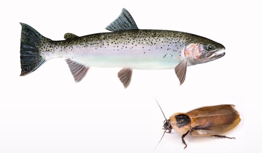
Aurora Scientific has helped thousands of laboratories perform functional, contractile testing of muscle for nearly 25 years. Though most of these laboratories work on murine models, we would like to recognize the outstanding research from a handful of labs who work on less common animal models and the incredible discoveries that have been made as a result. This publication review will highlight the use of two unconventional animal models for muscle physiology research: the cockroach (Blaberus discoidalis) and the steelhead trout (Oncorhynchus mykiss).
“TC. Tune et al. (2020) simultaneously measured the myofilament lattice spacing, packing structure and macroscopic force production of these two leg muscles to test whether structural differences in the myofilament lattice might correspond to the muscles’ different mechanical functions.”
Nanometer-scale structure differences in the myofilament lattice spacing of two cockroach leg muscles correspond to their different functions
The two muscles of a cockroach leg have almost identical physiological properties. However, during running, one muscle is bifunctional in that it produces positive mechanical work as well as a ‘braking’ function while the other acts only as a ‘brake’. Using time-resolved X-ray diffraction in intact, contracting muscle, TC. Tune et al. (2020) simultaneously measured the myofilament lattice spacing, packing structure and macroscopic force production of these two leg muscles to test whether structural differences in the myofilament lattice might correspond to the muscles’ different mechanical functions. While the structural patterns are very similar, one muscle has 1 nm smaller lattice spacing than the other while in a resting position.
Under isometric stimulation using our 305C Dual-Mode Muscle Lever System, the difference in lattice spacing disappears, which is consistent with the two muscles’ identical steady-state behavior. During periodic contractions, one muscle undergoes a 1 nm increase in lattice spacing, which correlates with greater force . This is the first identified structural feature in the myofilament lattice of these two muscles that shares their whole-muscle dynamic differences and quasi-static similarities.
“To examine if this diminished cardiac function may be related to compromised myocardial contractility, C. Carnevale et al. (2020) used our 300C-LR Lever System and our 404A Force Transducer to measure work and power in spongy myocardial strips from normoxic- and hypoxic-acclimated steelhead trout.”
Hypoxic acclimation negatively impacts the contractility of steelhead trout (Oncorhynchus mykiss) spongy myocardium
Cardiac function is diminished in Atlantic cod and rainbow trout following acclimation to hypoxia at 10–12°C. Based on past studies, this compromised cardiac stroke volume is not due to structural changes in the heart of these fish. To examine if this diminished cardiac function may be related to compromised myocardial contractility, C. Carnevale et al. (2020) used our 300C-LR Lever System and our 404A Force Transducer to measure work and power in spongy myocardial strips from normoxic- and hypoxic-acclimated steelhead trout. Work required to lengthen the muscle, as during filling of the heart, was strongly dependent on frequency but was not affected by hypoxic acclimation or test PO2. In addition, strips from hypoxic-acclimated trout recovered less net power when returned to normoxia.
These results strongly suggest that acclimation to hypoxic environments reduces myocardial contractility, and in turn, may limit stroke volume, but that this diminished performance does not improve the capacity to maintain myocardial performance under oxygen-limiting conditions. Understanding of the relationship between water O2 levels and myocardial function in these fish, and the mechanisms involved in the cardiovascular compensation for their environment, is key to understanding how fish populations will respond to climate change.


