As we close out the first quarter of the century and venture into 2026, the start of the new year brings refreshed hopes, forward-looking resolutions, and aptly enough, a focus on pigs. In German-speaking countries, pigs have long symbolized good luck and prosperity. Glücksschwein, whether marzipan, ceramic, or even decorated lemons with coin tails, are gifted to bring fortune, reflecting the idea that pigs root forward into progress. The German phrase Schwein haben—“to have a pig”—means to be lucky, while traditional pork dishes on New Year’s Day are thought to ensure abundance. It’s a fitting metaphor for this month’s publication review on porcine research. Recent studies using pigs and swine-derived tissues are uncovering muscle and cardiac mechanics, offering a lucky start to 2026.
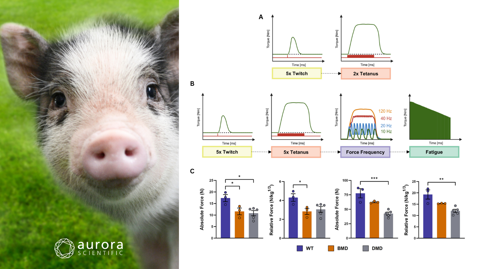
Featured image (photo of pig by HadelProductions from Getty Images Signature, via Canva.com with figures adapted from ©Blasi et al. (2025), licensed under CC BY 4.0) shows the experimental protocol and peak force measurements. A) preliminary twitch and tetanic stimulations used to verify electrode placement and stimulation parameters. B) the main data collection, including multiple twitch and tetanic stimulations, force–frequency measurements, and a 10-minute fatigue protocol to assess torque decline over time. C) the resulting peak twitch forces, both as absolute values and normalized to body mass, highlighting differences between DMD, BMD, and WT pigs with statistical significance indicated (*p < 0.05; **p < 0.01; ***p < 0.001).
Reduced Muscle Force in Dystrophic DMDΔ52 Pigs Is Incompletely Restored by Systemic Transcript Reframing (DMDΔ51–52)
Duchenne muscular dystrophy (DMD) is a devastating neuromuscular disorder marked by dystrophin deficiency and progressive loss of muscle strength. Over the years, large animal models have increasingly been used to bridge the gap between preclinical studies and human disease. In fact, pigs lacking DMD exon 52 recapitulate severe functional and pathological features of DMD, while transcript reframing via additional deletion of exon 51 restores dystrophin expression and ameliorates disease hallmarks. However, whether this molecular correction translates into full recovery of intrinsic skeletal muscle force has remained unclear. Therefore, Blasi et al. (2025) quantitatively assessed in-vivo muscle function in dystrophic and transcript-reframed pigs to determine the extent to which systemic exon skipping restores muscle performance.
To assess skeletal muscle dysfunction and its restoration in dystrophic pigs, age-matched DMD (DMDΔ52), BMD (DMDΔ51–52), and wild-type (WT) littermates were generated through targeted breeding and genotyping. In-vivo muscle function was quantified under general anaesthesia using Aurora Scientific’s 892A Swine Isometric Footplate Test Apparatus. Specifically, dorsiflexor muscle torque was measured in response to controlled twitch, tetanic, force–frequency, and fatigue stimulation protocols. Complementary analyses included serum clinical chemistry, detailed histopathology, immunohistochemistry and immunofluorescence, and quantitative proteomics.
Across genetically matched cohorts, DMD pigs exhibited pronounced muscle pathology, altered fibre composition, and elevated serum markers of muscle damage, whereas BMD pigs largely resembled WT animals at the molecular and histological levels. Neural-evoked in-vivo measurements revealed substantially reduced twitch and tetanic forces in DMD muscle, with BMD pigs showing partial but incomplete recovery of peak force and contraction dynamics relative to WT controls. Beyond strength deficits, DMD muscle displayed markedly slowed contraction and relaxation kinetics, and an earlier onset of fatigue, while BMD muscle demonstrated near-normal timing properties despite residual force limitations. Collectively, these findings demonstrate that systemic transcript reframing restores dystrophin expression and ameliorates many structural and functional deficits. However, it does not completely normalize skeletal muscle performance, underscoring the value of quantitative force measurements for therapeutic evaluations in large-animal DMD models.
Myosin Modulator Aficamten Inhibits Force by Altering Myosin’s Biochemical Activity Without Changing Thick Filament Structure
Hypertrophic cardiomyopathy has emerged as a paradigmatic disease of sarcomeric hypercontractility, driving interest in small-molecule myosin inhibitors that directly modulate cardiac force generation. While the clinical efficacy of agents such as mavacamten is well established, the precise mechanisms by which newer compounds like aficamten regulate myosin activity within intact sarcomeres are unknown. In particular, it has been unclear whether aficamten suppresses force by structurally sequestering myosin heads or by altering the biochemical kinetics of the motor itself. Mohran et al. (2026) addressed this gap by integrating structural, biochemical, and mechanical assays to define how aficamten modulates myosin ATPase activity and contractile behaviour without disrupting thick filament organization.
To define the mechanisms by which aficamten modulates cardiac myosin function, porcine myocardium and human iPSC-derived engineered heart tissues were employed. Isometric force production and cross-bridge kinetics were assessed in permeabilized left ventricular strips mounted between Aurora Scientific’s 400A force transducer and 315C High-Speed Length Controller. This enabled precise measurements of calcium-dependent force generation and rates of tension redevelopment under controlled sarcomere lengths. It also provided high-resolution quantification of myosin-driven mechanics in the presence of aficamten or mavacamten, directly linking biochemical modulation to contractile output. Further analyses included single-molecule ATP turnover imaging, x-ray diffraction to assess thick filament structure, sinusoidal length-perturbation analysis, and intact twitch measurements in engineered heart tissues to integrate molecular effects across multiple functional scales.
Throughout the experimental scales, aficamten consistently reduced cardiac force generation by shifting myosin into slower biochemical ATPase states without inducing the structural sequestration of myosin heads along the thick filament. Measurements from permeabilized porcine myocardium revealed a marked reduction in maximal calcium-activated force and calcium sensitivity, while rates of tension redevelopment and cross-bridge cycling kinetics remained largely unchanged. At the myofibril and engineered heart tissue levels, aficamten further accelerated relaxation kinetics and reduced twitch tension, distinguishing its functional profile from mavacamten despite comparable force inhibition. Together, these results demonstrate that aficamten suppresses contractility primarily through biochemical modulation of myosin activity rather than structural reorganization. This finding provides critical mechanistic insight into how next-generation myosin inhibitors fine-tune cardiac performance in hypertrophic cardiomyopathy.
ATP directly modulates thick filament structure and function in porcine myocardium
Cardiac contraction is fundamentally constrained by energy availability, as ATP hydrolysis by myosin powers the cross-bridge cycle that sustains cardiac output. Although reduced myocardial ATP levels are a well-documented feature of heart failure and other cardiomyopathies, prior work has largely suggested that actomyosin ATPase activity is insensitive to ATP concentration within the physiological millimolar range. Consequentially, the mechanistic consequences of ATP depletion remain unresolved. This gap has obscured how energetic failure translates into impaired contractility and relaxation at the level of the sarcomere. For this reason, Rhodehamel et al. (2025) tested whether ATP itself modulates thick filament structure and mechanics in permeabilized porcine myocardium, thereby linking metabolic state to myosin activation and cardiac function.
To examine how ATP directly regulates thick filament structure and cardiac mechanics, permeabilized porcine left ventricular myocardium were used, along with isolated cardiomyocytes subjected to controlled biochemical and mechanical manipulations. Structural changes in myosin organization were assessed using small-angle x-ray diffraction, with muscle strips mounted to Aurora Scientific’s 402B Force Transducer to precisely set sarcomere length and monitor passive tension during ATP titrations. Functional contractility was quantified using our force and length control systems (403A: Force Transducer, 315C: High-Speed Length Controller, and 600A: Real-Time Muscle Data Acquisition and Analysis System) to measure isometric tension–calcium relationships, cross-bridge attachment and detachment kinetics, and force–velocity–power output under varying ATP concentrations.
Increasing ATP concentration in permeabilized porcine myocardium caused structural rearrangements in myosin, as evidenced by decreased intensities of the M3 meridional reflection and the first myosin layer line (IMLL1). This, along with increased interfilament lattice spacing (d1,0) and I1,1/I1,0 ratios, indicated myosin heads move radially toward actin. X-ray diffraction also showed that ATP promotes a partial OFF-to-ON transition of myosin heads, with increases in SM6 and reductions in IM3 and IMLL1, suggesting a distinct ATP-mediated mechanism compared with calcium-induced activation. Functional measurements revealed that higher ATP increased cross-bridge attachment (2πb) and detachment (2πc) rates, shifted the tension–calcium relationship upward at submaximal calcium, and enhanced both maximal shortening velocity (Vmax) and peak power (Pmax) in cardiomyocytes. Together, these results demonstrate that ATP directly modulates both the structural activation of thick filaments and the mechanical performance of cardiac muscle, linking metabolic state to contractile function.
Conclusions
These studies by Blasi et al. (2025), Mohran et al. (2026), and Rhodehamel et al. (2025) illuminate how porcine models can reveal the structural, biochemical, and functional underpinnings of muscle and cardiac performance. Collectively, they underscore the power of pigs as translational models and bring a pig-ture perfect measure of scientific good fortune to the year ahead.