As we step into February, the month dedicated to matters of the heart, there’s no better time to reflect on the latest strides in cardiac research. Whether you’re nostalgic for Valentine’s Day or simply embracing Heart Month, this time of year reminds us of the importance of a healthy heart and the groundbreaking science devoted to its understanding. Recently, researchers have made heart-hitting discoveries, including a novel method to assess heart tissue integrity, the intricate relationships between heart function and redox balance, and the effects of chemotherapy on cardiorespiratory muscle health.
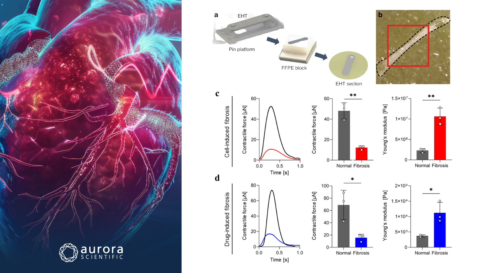
Featured image (photo by SDenisov from Getty Images, via Canva.com with figures adapted from ©Park et al. (2026), licensed under CC BY 4.0) depicts the preparation and mechanical characterization of engineered heart tissues (EHTs). A) preparation of an EHT section, B) picture of the EHT slide, and C-D) mechanical characterization of fibrotic EHTs (n = 3, mean ± standard deviation, *p < 0.05, **p < 0.01).
Label-free mid-infrared dichroism-sensitive photoacoustic microscopy for histostructural analysis of engineered heart tissues
Biological tissues such as cardiac muscle and tendons are known for their highly organized microstructure, which is critical to their physiological function. Current histological techniques for analyzing tissue alignment are often labor-intensive, antibody-dependent, and lack objective measurement. To address this, Park et al. (2026) introduced label-free mid-infrared dichroism-sensitive photoacoustic microscopy (MIR-DS-PAM) as a novel, efficient method to assess the microstructural integrity of engineered heart tissues (EHT).
The MIR-DS-PAM system used a nanosecond pulsed quantum cascade laser with controlled polarization and power monitoring to focus mid-infrared light onto the EHT sample, generating photoacoustic signals detected by a 30 MHz ultrasound transducer. The amplified and filtered signals were then digitized and recorded while motorized stages enabled scanning, with 250 A-lines averaged per pixel at an average laser power of ~0.26 mW. To evaluate the mechanical properties of the EHTs, Aurora Scientific’s 1500A: Isolated Muscle System was employed, enabling precise force measurements and calculation of Young’s modulus under standardized conditions. Additional methods included immunofluorescence staining for tissue characterization, and action potential propagation assessments to analyze electrophysiological properties.
Measurements in fibrotic EHTs revealed a significant reduction in force and an increase in Young’s modulus in both cell-induced and drug-induced fibrosis models, indicating mechanical dysfunction. MIR-DS-PAM provided detailed insights into the alignment and organization of the extracellular matrix (ECM) in both normal and fibrotic tissues, showing a disruption in ECM structure in fibrotic EHTs. These findings underscore the utility of MIR-DS-PAM in capturing detailed histostructural and mechanical characteristics of EHTs. By combining molecular specificity with polarization sensitivity, MIR-DS-PAM offers a more reliable and objective approach to quantifying tissue alignment, which is key to evaluating both tissue development and conditions like fibrosis.
Modulation of cardiac contractility and redox balance via cannabinoid type II receptor activation in healthy rats
The endocannabinoid system, particularly the cannabinoid receptor type II (CB2r), has been implicated in various cardioprotective actions, including anti-inflammatory and antioxidant effects, in pathological models of heart disease. However, the effects of CB2r activation on healthy myocardial tissue remain largely unexplored. Therefore, Fernandes et al. (2026) examined the impact of CB2r activation on cardiac contractility, calcium-handling proteins, and redox balance in healthy rats.
Male Wistar rats were randomly assigned to two treatment groups, receiving either a vehicle or the CB2r-selective agonist HU-308, and were euthanized 24 hours post-treatment. Cardiac function was assessed ex-vivo using left-ventricle myocardial strips in a thermostatically controlled bath, with Aurora Scientific’s 701C: Electrical Stimulator and an isometric force transducer. In particular, contractility measurements included force-frequency relationships and cardiac pumping capacity. For protein expression analysis, Western blotting was performed on myocardial samples to evaluate Ca2+-handling proteins, including SERCA2a, PLB, and NCX1. Additionally, redox biomarkers such as superoxide dismutase, catalase, and glutathione levels were measured to assess antioxidant activity and oxidative stress in the myocardium.
The HU-308 treatment group exhibited significant improvements in cardiac function, including increased force of contraction (Fc), contraction rate (+dF/dt), relaxation rate (−dF/dt), and cardiac pumping capacity (CPC), compared to vehicle-treated controls. This positive modulation of myocardial contractility was further supported by increased expression of key Ca2+-handling proteins such as SERCA2a, PLB, and NCX1, and a notable rise in cAMP levels. Contraction forces across different pacing frequencies revealed an 88% to 142% enhancement in contractility with HU-308 treatment. Additionally, CB2r activation improved antioxidant defenses, as evidenced by elevated SOD, GPx, and GR activity, along with reduced markers of oxidative damage, including lipid peroxidation, protein carbonylation, and DNA strand breaks. These results suggest that CB2r activation enhances both cardiac function and oxidative stress resistance, positioning it as a potential therapeutic strategy for improving myocardial health.
Sex Differences in Response to Acute Doxorubicin Cardiorespiratory Muscle Dysfunction and Preconditioning Exercise
Doxorubicin (DOX) is an effective chemotherapeutic agent, but its use is limited by cardiorespiratory muscle toxicity, leading to muscle dysfunction and exercise intolerance. While exercise has been shown to alleviate some of the negative effects of DOX, the impact of preconditioning exercise on male and female responses to DOX exposure remains unclear. For this reason, Montalvo et al. (2026) evaluated sex differences in the severity of DOX-induced muscle dysfunction and the protective effects of preconditioning exercise.
Male and female Sprague-Dawley rats were employed to explore the impact of preconditioning exercise and doxorubicin (DOX) on cardiorespiratory muscle function. Rats underwent a 2-week exercise protocol, followed by a saline or DOX injection, before undergoing various tests including exercise tolerance, plethysmography, and echocardiography. Diaphragm force production was assessed ex-vivo using Aurora Scientific’s 1205A: Isolated Muscle System for Rats, where diaphragm muscle strips were mounted in a tissue bath to measure specific force and fatigue. Additionally, muscle tissue was analyzed for fiber type and cross-sectional area, while gene expression and DOX metabolites were quantified through RNA sequencing and HPLC.
Exercise preconditioning increased endurance capacity in both male and female rats, including those treated with DOX, compared with their sedentary counterparts. Acute DOX administration reduced left ventricular fractional shortening and diaphragm maximal force production in both sexes; however, exercise preconditioning prevented these declines in cardiac and diaphragm function only in female rats, with no functional protection observed in males. Therefore, while DOX induced cardiorespiratory muscle toxicity in both sexes, the protective effects of exercise preconditioning on muscle function were sex-specific and limited to females. Overall, these results highlight the impact of DOX on muscle and cardiorespiratory function, emphasizing the complex interactions between exercise preconditioning, sex, and chemotherapy, which could inform strategies to mitigate muscle toxicity in cancer patients.
Conclusions
These studies by Park et al. (2026), Fernandes et al. (2026), and Montalvo et al. (2026) delve into critical areas of cardiac health, from innovative imaging techniques to cannabinoid-based therapies and the protective effects of exercise preconditioning against chemotherapy-induced damage. Together, they highlight the dynamic interplay between tissue integrity, redox balance, and muscle function, offering promising avenues for improving cardiovascular care.